 Home
Home
|
Table of Content - Volume 18 Issue 2 - May 2021
Functional outcomes of calcaneal fractures managed with external fixator application
Sunil Malhotra1, Mohd Shifa Hasan2, Shrikant Kashyap3*, Kamal Swarn4
1Associate Professor, 2Assistant Professor, 3Post Graduate Resident, 4Professor, Department of Orthopaedics, Subharti Medical College, Meerut, INDIA. Email: dr.shrikantkashyap@gmail.com
Abstract Background: The most common fracture of tarsal bones is calcaneum fractures. Fewer complications are associated with closed and percutaneous techniques as compared with open reduction techniques. Aims and objectives: The aim of the study was to analyse the functional outcome of calcaneal fractures treated by external fixator application. Methods: This prospective study conducted over duration of 2 years in orthopaedics department of Netaji Subhash Chandra Bose Subharti medical college, Meerut. It consists of 30 cases with displaced calcaneal fracture treated using percutaneous reduction technique followed with external-fixator application. Patients were followed up for minimum of 12 months. The functional outcome was evaluated using American Orthopaedic Foot and Ankle Society (AOFAS) - Hind foot score. Results: The mean age was 37.4 years and majority were male’s 17 cases (56.66%). The mean Bohler’s and Gissane’s angle preoperative was 12°, and 154.60° respectively which improved postoperatively and at final 12 months follow-up was 30.40° and 144.70° respectively. At final 12 months follow-up, the functional outcome calculated on basis of AOFAS score was excellent in 8 (26.6%), good in 20 (66.74%), fair in 1 (3.33%) and poor in 1 (3.33%). Mean AOFAS score was good (84.62). Complications were reported in four cases with pin-site infection. Conclusions: This method is effective and has good functional outcome with lesser complications. Keyword: Calcaneal fracture, external fixator, Bohler’s angle, Gissane’s angle.
INTRODUCTION Calcaneal fractures are the most common fractures of the tarsal bones. As per literature, initially these fractures were managed conservative which over the years evolved to surgical modalities. Although open reduction has been performed since the early 1930s, infection and technical problems lead surgeons against operative treatment. Overall, due to better imaging techniques, minimal invasive techniques and antibiotics improved the results of operative fracture fixation.1 In this study we tried to analyses functional outcomes of calcaneal fractures managed by percutaneous reduction and external fixator application. We also tried to evaluate if this management modality has any advantage over other prevailing modalities of management. AIMS AND OBJECTIVES To analyse the functional outcomes of external fixator application in management of fractures calcaneum. Advantage of external fixator over other modalities of management of calcaneus fractures.
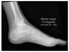
MATERIAL AND METHOD This prospective observational study was carried out on patients of intra-articular calcaneal fracture as per inclusion criteria in Department of Orthopaedic, N.S.C.B.S. Medical College, Meerut over a period of 2 years. A total 36 patients presented with calcaneum fracture out of which 3 were not fitting in our inclusion criteria while 3 refused to take part in the study. Finally, 30 patients were enrolled for the study which was followed up post operatively, at 1 month, 2months, 6 months and 12 months. Ethical approval was obtained for the study from the Ethics Committee of N.S.C.B.S. medical college and hospital, Meerut. Inclusion criteria: Skeletally mature patients of both genders presenting with unilateral displaced calcaneal fractures presenting within 2 weeks of injury. Exclusion criteria: bilateral calcaneus fractures, undisplaced and more than 2 weeks old fractures. Pre-operative evaluation: At the time of admission radiographs (with standard magnification) with antero-posterior, lateral, axial view of calcaneum of fractured and normal side were taken. From the radiographs, the type of fracture was determined, and the pre-operative Bohler’s angle2, Gissane’s angle3 was measured of both sides. Anti-oedema measures were taken such as elevation of limb, below knee plaster slab, antibiotics and daily dressing in cases associated wound and anti-inflammatory drugs. On lateral view of the radiograph of the calcaneum is used to identify Bohler’s angle usually 20 to 40 degree, decreases in this angle indicates the weight bearing surface of the calcaneum has collapsed and shifting the body weight anteriorly.2 It is formed by drawing two lines first line starts from the highest point on the anterior process of the calcaneum to highest point on the posterior facet and the second line drawn tangential to the superior edge of the tuberosity (figure 1).
Figure 1: Bohler’s angle The critical angle of gissane3 the first line extends along the lateral border of the posterior facet, the second extends anteriorly to the beak of the calcaneum (figure 2).
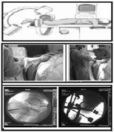
Figure 2: Gissane’s angle Operative Technique:4 The patient was placed in prone position on a radiolucent operating table, with the foot protruding out of the operating table. Kirschner wires (3mm) were inserted from the medial side through the calcaneal tuberosity and the talar neck respectively. A third K-wire was inserted into the cuboid bone if required. The direction of the Kirschner wires was chosen on the basis of the fracture displacement. Pointed reduction forceps were used to squeeze the inner and outer wall of calcaneus, restoring the width of the calcaneus and further correction were done using blunt drifter (punch) was inserted to unlock the depressed part in order to restore joint congruence if needed (figure3). Joshi’s external stabilization system were applied on each side and widened apart to restore calcaneal height and length. Final reduction was checked under image intensifier in AP and axial views. A compression dressing was applied on the operated site after surgery.
Figure 3: Intraoperative technique clinical and c-arm imaging Postoperative protocol:
Follow up protocol: •All patients were followed up at interval of 1 month, 2months, 6 months and 12 months. •The functional outcome was evaluated in accordance with The American Orthopaedic Foot and Ankle Society (AOFAS) - Hind foot score5.
OBSERVATIONS AND RESULTS Mean age of the patient at the time of presentation was 37.4 years (range 19 to 52 years). Majority of them were male 17 cases (56.66%) and 13 cases (43.34%) were females. Injury due to fall from height predominated the series by 60% (n=18) followed by road traffic accident 40% (n=12). All cases were assessed radiologically pre-operatively and were classified using Essex-Lopresti classification6. The incidence of joint depression type (56.66%; n=17) was more common than tongue type (43.34%; n=13) and comminuted type (10%; n=3). 10% (n=3) of the cases presented with associated injuries (spinal injury, pelvic fractures, and multiple fractures). Radiological assessment: A. Bohler’s angle: The mean was 12° pre-operatively which was restored to mean 33.50° as of post-operative day 1 (figure 4). At the final 12 months follow up mean Bohler’s angle was 30.40° when compared to contra lateral normal calcaneus with mean Bohler’s angle of 36.50°, the difference value was 6.1° (table 1). B. Gissane’s angle: The mean Gissane’s angle prior to operative intervention was 154.60° which was restored to mean 141.50° immediate post-operative (day 1). At the final 12 months follow up mean Gissane’s angle was 144.70° (figure 4) when compared to contra lateral calcaneus with mean Gissane’s angle of 137.80° the difference value was 6.9° (table 1).
Table 1: Mean Bohler’s and Gissane’s angle on radiological assessment
Figure 4: Radiograph showing Bohler’s and Gissane’s angle. a. Preoperative b. Immediate post-operative day 1 c. at 1 month follow-up d. at 12 months follow-up Functional outcome The mean American Orthopaedic Foot and Ankle Society (AOFAS)- Hind foot score (table 2) was 62.30 at 2 months and was found to be better at 6 months 87.50 following which there was a slight decrease in the mean score at 12 months (84.62). Table 2: American Orthopaedic Foot and Ankle Society (AOFAS) - Hind foot mean score at follow ups
Complications There were 4 cases (13.34%) with complications out of the 30 cases. All the four cases were of superficial pin site infections that responded to infiltration of local antibiotics and oral antibiotics. These cases had less AOFAS score and more deterioration in radiological parameters compared to other cases. Fracture union was observed in all the 30 cases (100%) on radiological assessment. The average time for union (radiological) was 10.4 weeks ranging from 8 to 12 weeks. Average time for the external fixator removal was 12.4 weeks.
DISCUSSION Essex-Lopresti classification was used in this study to identify the fracture pattern. The majority of cases were categorized under joint depression type (56.66%; n=17) was more common than tongue type (43.34%; n=13). This was in concordance with the studies by Magnan B., et al. (2006)7 The results of AOFAS - Hind foot score in this study are better to that observed by Singh A., et al. (2008)8 they used minimally invasive techniques in their studies (Table: 4). When compared to another study conducted by Siebe D.B., et al.9 (2015), the average AOFAS - scores at 12 months in this study was better to both the non-operatively treated group and the open reduction internal fixation group (table 3).
Table 3: Functional outcome assessment using AOFAS- hind foot Score in different studies
When compared to another study conducted by Kissel C.S., et al.10 (2011), Bhavik Y.D., et al. (2016)11 and Jain S., et al. (2013)12 the mean Bohler’s and Gissane’s angle at 12 months in this study was better to both the non-operatively treated group and the open reduction internal fixation group (table 4).
Table 4: Radiological parameters average in different studies
In this study, there were 4 cases (13.34%) with complications out of the 30 cases. The overall complication rate in this study was less when compared to other studies (table 5).
Table 5: Complication with various method of management in different studies
CONCULSION External fixation technique has proven to be safe and can be applied in calcaneal fractures when surgery is indicated. The technique restores the Bohler’s angle and Gissane’s angle and has a good functional outcome making it a viable alternative for the treatment of fractures of the calcaneus. Limitations of this study are the small number of patients and no direct comparison with another method of treatment. The disadvantage is possibility of achieving incomplete reduction of fracture. The study did not include CT-Scan due to financial restrains.
REFERENCES
Policy for Articles with Open Access: Authors who publish with MedPulse International Journal of Pediatrics (Print ISSN: 2579-0897) (Online ISSN: 2636-4662) agree to the following terms: Authors retain copyright and grant the journal right of first publication with the work simultaneously licensed under a Creative Commons Attribution License that allows others to share the work with an acknowledgement of the work's authorship and initial publication in this journal. Authors are permitted and encouraged to post links to their work online (e.g., in institutional repositories or on their website) prior to and during the submission process, as it can lead to productive exchanges, as well as earlier and greater citation of published work.
|
|
|||||||||||||||||||||||||||||||||||||||||||||||||||||||||||||||||||||||||||||||||||||||||||||||||||||||||||||||||||||||||||||||||||||||||||||||||||||


 This work is licensed under a Creative Commons Attribution-NonCommercial 4.0 International License.