 Home
Home
|
Table of Content - Volume 20 Issue 2 - November 2021
Sagittal alignment Functional and Neurological outcome with skipped level instrumentation with lateral mass screw for multilevel Cervical Myelopathy with OPLL
Anil kumar D N
Associate Professor, Department of Orthopaedics, Ayaan Institute of Medical Sciences, Hyderabad, INDIA. Email: anildn@gmail.com
Abstract Background: Posterior cervical laminectomy and instrumentation is a preferred procedure to treat multilevel cervical myelopathy with OPLL. In this study, we evaluated effect of skipped levels of instrumentation with posterior decompression by laminectomy on post op sagittal alignment, functional and neurological outcome in cervical spondylotic myelopathy patient with OPLL. Materials and Methods: In this retrospective study conducted between June 2003 and December 2016, we have evaluated 19 patients with multilevel cervical myelopathy with OPLL who underwent multilevel cervical laminectomy and instrumentation with lateral mass screw. All patients were evaluated preoperatively and postoperatively with Nurick’s grading and Modified Japanese Orthopaedic Association (mJOA) scale for neurological function. Cooper scale and British Medical Research Council grading system for motor function. Alignment of the cervical spine was done preoperatively and postoperatively by calculating the curvature index. Axial MRI was used to calculate the severity of compression preoperatively which was calculated as per Singh’s criteria and postoperatively to assess the adequacy of decompression at the operated level. Results: In this study, there were 19 patients including 9 males and 10 females, with mean age of 58.3 years the mean duration of follow up of patients was 29.24 months. In total, cervical laminectomy was performed at 59 levels in 19 patients with an average of 3 level laminectomies, and in total, 41 lateral mass screws were inserted. On postoperative follow up the mJOA and Nurick’s grading showed improvement in all cases as compared to preoperative findings. The mean Cooper scale also showed significant improvement in both upper and lower limbs postoperatively (P < 0.001). The mean preoperative Cooper scale was 1.75 and postoperative was 0.31 for upper limbs, and the mean Cooper scale was 2.14 preoperatively and 0.56 postoperatively for lower limbs. X-rays done on routine follow ups showed good alignment of the cervical spine with maintenance of curvature index in all patients. Conclusion: The multilevel cervical laminectomy and skipped level instrumentation with lateral mass screw for multilevel cervical myelopathy with OPLL is a safe and cost-effective technique that provides decompression of the spinal cord, prevents the development of kyphotic spinal deformity by maintaining posterior tension band
INTRODUCTION Cervical spondylosis secondary to degeneration of intervertebral disc, facet joints, posterior longitudinal ligament, ligamentum flavum, and ossification of posterior longitudinal ligament (OPLL)are the most common causes of cervical myelopathy which can lead to irreversible neurological impairment.1-3 The standard surgical treatment for cervical myelopathy consists of either anterior decompressive procedure or posterior decompressive procedure. Anterior decompressive procedure consists of anterior discectomy and fusion or corpectomy and fusion.4,5 Posterior decompressive procedure consists of either laminoplasty or laminectomy with or without instrumentation.6-8 The pathophysiology behind cervical myelopathy is direct compression of the cord and ischemic insult to the cord as a result of reduced blood flow. Anterior cervical decompression surgery helps to remove the direct compression on the cord as well as helps to increase the blood flow to the spinal cord. Anterior cervical discectomy and fusion is gold standard for single-level cervical myelopathy and has shown to produce good clinical results.9 However, in patients with multiple segment involvement with myelopathy, the approach toward the treatment is unclear.8 Multilevel anterior surgery is associated with complications such as increased surgical trauma and increased incidence of pseudarthrosis, graft dislodgement, and implant failure as the number of level increases. It is also associated with increased incidence of adjacent segment degeneration and neurological deterioration.1 Multilevel posterior surgeries such as laminoplasty or laminectomy without instrumentation are associated with complications such as instability, kyphosis, axial pain, perineural adhesion, neurological deterioration, and C5 nerve root palsy. However, multilevel cervical laminectomy with lateral mass screw fixation provides immediate stability, hence prevents the development of kyphotic deformity and adjacent segment degeneration by the prevention of osteophyte formation. The goal in treating multilevel cervical myelopathy is to achieve adequate decompression without compromising the stability of the cervical spine. Posterior cervical laminectomy and instrumentation is a preferred procedure to treat multilevel cervical myelopathy with OPLL. In this study, we evaluated effect of skipped levels of instrumentation with posterior decompression by laminectomy on post op sagittal alignment, functional and neurological outcome in cervical spondylotic myelopathy patient with OPLL.
MATERIALS AND METHODS In this retrospective study conducted between October 2019 and October 2021in Ayaan institute of medical sciences Hyderabad, we have evaluated 19 patients with multilevel cervical myelopathy with OPLL who underwent multilevel cervical laminectomy and instrumentation with lateral mass screw. All patients were evaluated preoperatively and postoperatively with Nurick’s grading and Modified Japanese Orthopaedic Association (mJOA) scale for neurological function. Cooper scale and British Medical Research Council grading system for motor function. Alignment of the cervical spine was done preoperatively and postoperatively by calculating the curvature index. Axial MRI was used to calculate the severity of compression preoperatively which was calculated as per Singh’s criteria and postoperatively to assess the adequacy of decompression at the operated level
RESULTS In this study, there were 19 patients including 9 males and 10 females, with mean age of 58.3 years the mean duration of follow up of patients was 13.24 months. In total, cervical laminectomy was performed at 59 levels in 19 patients with an average of 2.15 laminectomies, and in total, 41 lateral mass screws were inserted. On postoperative follow up the mJOA and Nurick’s grading showed improvement in all cases as compared to preoperative findings. The mean Cooper scale also showed significant improvement in both upper and lower limbs postoperatively (P < 0.001). The mean preoperative Cooper scale was 1.75 and postoperative was 0.31 for upper limbs, and the mean Cooper scale was 2.14 preoperatively and 0.56 postoperatively for lower limbs. X-rays done on routine follow ups showed good alignment of the cervical spine with maintenance of curvature index in all patients.
Table 1: Nurick’s grading
Table 2: mJOI= Modified Japanese orthopaedic Association
Table 3: COOPER SCALE
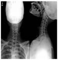
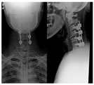
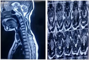
Figure 1: Pre op x ray Figure 2: Pre op MRI I Figure 3: POST OP X RAY Figure 4: POST OP MRI
DISCUSSION Multilevel cervical myelopathy can be treated with either anterior or posterior decompressive procedure; the choice of surgery depends on the location of pathology, alignment of the cervical spine, and also surgeon’s preference.25,26 In this study, we have evaluated the outcome of multilevel posterior cervical laminectomy with instrumented fusion with lateral mass screw. The results of our study show that patients with multilevel cervical myelopathy operated with this technique have favorable neurological and functional outcome as measured by mJOA score, Cooper scale, and Nurick’s grading. The reason for favorable result is good surgical exposure, excellent cord decompression, accurate placement of the screw in lateral mass, good bone graft for fusion, and avoidance of screw penetration in the spinal canal or disc space. This factor helps in the maintenance of cervical spine alignment and prevents the development of cervical kyphosis as the loss of alignment of cervical spine and development of cervical kyphosis may affect the outcome after surgery. Maintenance of alignment and adequate decompression are the most important factors in achieving good clinical outcome as micromotion in degenerated cervical spine can lead to continuous irritation of the already compromised cord and can prevent the progress of neurological recovery.23,27 Laminoplasty or laminectomy without instrumentation for multilevel pathology is associated with complications such as neurological deterioration and progressive kyphotic deformity. As the posterior neck muscles provide tension band, it keeps the cervical spine maintained in lordosis, as these muscles are detached and facet joints are removed during laminoplasty and laminectomy which undergoes significant atrophy causing loss of cervical lordosis and leading to progressive kyphotic deformity.28 Facetectomy done while doing laminectomy is also one of the important causes of postoperative kyphosis after laminectomy.29 Excluding patients with kyphotic cervical spine helps toprevent the bowstringing of the decompressed cervical spinal cord over the anterior osteophytes or vertebra body.9,21,23 Postoperative MRI done in our study showed that the cervical cord moved backward away from the anterior osteophytes after posterior decompression, providing indirect decompression of the cervical spinal cord and helps in good postoperative outcome. The results of our study can be compared with similar studies done in the past. In a similar study conducted by Kumar et al.,30 they concluded that, after cervical laminectomy and fusion for cervical myelopathy, 80% of patients had good clinical outcome and 76% of patients showed improvement in myelopathy scores. In their study, none of the patients had worsening of neurological symptoms, instability, or progression of kyphosis. They also stated that patients with better preoperative neurological status were likely to improve more as compared to patients with poor neurological status. In a study conducted by Chang et al.8 on 58 patients with multilevel cervical myelopathy who underwent cervical laminectomy and fusion with lateral mass screw, mJOA scores improved significantly in 85.5%, while 14.5% of patients showed no improvement. None of the patients had deterioration of mJOA score in their study. All patients showed radiographic fusion in dynamic X-ray done at an average followup duration of 11.9 months. Four patients had C5 nerve root palsy and one patient had superficial wound infection which settled without any sequelae. Huang et al.31 retrospectively studied 32 patients who were treated with cervical laminectomy and fusion with lateral mass screw for cervical myelopathy. The patients were evaluated by Nurick’s grading for clinical evaluation, and X-ray and MRI were done for radiological assessment both preoperatively and postoperatively in their study. Nurick’s grading showed significant improvement in 22 patients, 9 patients showed no improvement. However, none of the patients had worsening of Nurick’s grading. Postoperative MRI showed compression in one patient and myelomalacic change in 15 patients which was same in preoperative MRI. However, these patients had significant neurological recovery similar to patients who did not show compression or myelomalacic changes. One patient had pseudarthrosis, three patients had wound infection which required reoperation, and two patients had C5 nerve root palsy which settled gradually without any intervention. Houten and Cooper23 studied 38 patients with cervical myelopathy who underwent laminectomy and instrumentation with lateral mass fixation. The patients were evaluated clinically with mJOA score, Cooper scale,and 5-point muscle grading. X-ray and MRI were done both preoperatively and postoperatively for the assessment of cervical spine alignment and adequacy of decompression. Clinically significant improvement was seen in 97% of patients with mJOA scale improved to 15.8 from 12.9. Cooper scale also showed significant improvement from 1.8 to 0.7 for upper extremities and 1.0–0.4 for lower extremities. X-rays done at mean followup of 5.9 months showed no change in the alignment of cervical spine. Postoperative MRI showed significant improvement in compression grading from 2.46 to 0.16. They concluded that multilevel cervical laminectomy with instrumentation is an effective procedure with minimum morbidity, adequate cord decompression, and provides immediate stability. The authors also concluded that neurological outcome was similar to anterior procedures and avoided the complications associated with anterior procedures and multilevel laminectomy without instrumentation. Multilevel cervical laminectomy is associated with C5 nerve root palsy with incidence of 4.6% in spondylotic myelopathy and 8.3% in OPLL.21 Posterior shift of the spinal cord following laminectomy leads to the tethering effect on the nerve root resulting in C5 palsy.31 Incomplete removal of osteophytes and soft tissue from the neural foramina during decompression can also reduce the mobility of the nerve root and increase the risk of nerve root palsy. Reconstruction of the cervical spine following decompression in excessive lordosis can cause impingement of the C5 nerve root leading to palsy.32 In our study, none of the patients developed C5 nerve root palsy as the decompression was adequate with removal of osteophytes from the neural foramen and also undermining of the facet joint was done which provided enough space for dorsal shift of the nerve root. Postoperative epidural hematoma can occur as a result of bleeding from the edges of the bone from the facet joint while decompressing the neural foramen.21 Hemostasis must be achieved carefully with bipolar electrocautery and gelatin sponge. Postoperative epidural hematoma can lead to progressive neurological deficit. The neurological deficit is temporary if drained within time. The neurological deficit can be permanent, irreversible, and can also lead to death if the hematoma is not drained in time.33-35 In our study, two patients had postoperative epidural hematoma with transient paralysis. The hematoma was drained immediately and meticulous hemostasis was achieved. None of the patients had residual paralysis. Posterior cervical spine instrumentation with lateral mass screw is the most commonly used technique for subaxial stabilization of the cervical spine. The common complications associated with lateral mass screw are due to breach in the lateral mass leading to screw penetration in the ventral soft tissue which can cause injury to the vertebral artery, nerve roots, and the cervical cord.7 The accurate placement of the lateral mass screw is dependent on the surgeon’s experience. Adequate bony fusion is necessary to prevent the screw back out and rod breakage. In our study, none of the patients had complications related to lateral mass screw. The incidence of complication was low in our study because of appropriate case selection, adequate decompression of the spinal cord, meticulous hemostasis, accurate trajectory of the lateral mass screw, and restoration of the cervical spine in lordosis. In our study, age, sex, previous medical illness, duration of symptoms, degree of compression, and signal intensity changes on MRI were not found to affect the final outcome of surgical intervention. There are certain limitations of our study. This study did not have any control group, hence whether this study leads to better neurological outcome as compared to anterior surgery could not be studied. In addition, there was no comparison group to study the cervical alignment postoperatively, in which lateral mass screws were put at all the levels being decompressed. Since there was no deterioration in the alignment of the followup X-rays, we conclude that instrumentation at all levels is not mandatory in multilevel cervical myelopathy.
CONCLUSION The multilevel cervical laminectomy and skipped level instrumentation with lateral mass screw for multilevel cervical myelopathy with OPLL is a safe technique that provides decompression of the spinal cord, prevents the development of kyphotic spinal deformity by maintaining posterior tension band.
REFERENCES
Policy for Articles with Open Access
|
|

 This work is licensed under a Creative Commons Attribution-NonCommercial 4.0 International License.