 Home
HomeOfficial Journals By StatPerson Publication
|
Table of Content - Volume 3 Issue 3 - September2017
Distribution of various fractures observed in adults in tertiary care institute
Dnynesh Dattatrey Patil1, Narendra Maganlal Shirsat2*
1,2Associate Professor, Department of Orthopedics, Dr. Ulhas Patil Medical College and Hospital, Jalgaon, Maharashtra, INDIA. Email: patildnyanesh@yahoo.com
Abstract Background: A bone fracture is a medical condition in which there is damage in the continuity of the bone. A bone fracture can be the result of high force impact or stress or a minimal trauma injury as a result of certain medical conditions that weaken the bones, such as osteoporosis, bone cancer, or osteogenesis imperfecta, where the fracture is then properly termed a pathologic fracture. Aims and objectives: To study the distribution of various fractures observed in adults the study institution and their age and sex wise distribution. Materials and method: The present study was conducted in the department orthopedics of Dr Ulhas Patil Medical College and Hospital, Jalgaon. We searched the medical record of the hospital of the year 2014 and all the cases of fractures in adults were selected for the study. Only newly diagnosed cases were enrolled in the study. Patients with soft-tissue injuries and dislocations were excluded. The detail information of all the selected patients was retrieved from the computerized case records and noted on a prestructured proforma. The details included the demographic information of patients including age, sex etc. was retrieved from the records. Results: Total 1590 cases of various fractures were observed in the institute during the study year. Tibia (29.94%) was the most commonly fractures bone followed by femur (23.08%) and forearm (radius and or ulna) (14.97%). It was seen that the proportion of fracture was more in male in younger age and the proportion was increasing in female as the age advances. Sexwise distribution showed that fracture of femur, forearm and tibia were common among males whereas fracture of hip, spine and humerus were common in females. The agewise distribution of fracture showed that fracture of long bones was common in young individuals. Fracture of femur, forearm, humerus and tibia were common in the age group of 21 to 40 years of age. The proportion of fracture femur and humerus were also seen common in elderly individuals also. Conclusion: Thus we conclude that fracture of tibia, femur and forearm (radius and or ulna) were common in adults. The proportion of fracture was more in male in younger age and the proportion was increasing in female as the age advances. Key Words: Fracture, incidence, sex, age.
A bone fracture is a medical condition in which there is damage in the continuity of the bone. A bone fracture can be the result of high force impact or stress or a minimal trauma injury as a result of certain medical conditions that weaken the bones, such as osteoporosis, bone cancer, or osteogenesis imperfecta, where the fracture is then properly termed a pathologic fracture.1 Injury-related bone fractures contribute to an increase in morbidity, death, disability, and health expenditures across the age span. The incidence of bone fracture is impacted by many factors including age, race, gender, biology, physiology, body habitus, environmental exposure to fracture-producing injury mechanisms and access to prevention programs.2-6 Astley Cooper recognized the effects of ageing on the skeleton,7 and Bruns8 discussed the influence of age and gender on the incidence of various types of fracture. In recent year’s fractures, particularly those occurring in osteoporotic bone have become a major health issue. They are relatively common and treatment has become increasingly expensive and complicated. Despite this, there is little known about their epidemiology. Osteoporotic fractures represent an enormous public health burden. Worldwide, there were an estimated 1.66 million hip fractures in 1990, about 1,197,000 in women and 463,000 in men.9
MATERIALS AND METHOD The present study was conducted in the department orthopedics of Dr Ulhas Patil Medical College and Hospital, Jalgaon. The aim of the study was to study the distribution of various fractures observed in adults the study institution and their age and sex wise distribution. Thus for the purpose of study we searched the medical record of the hospital of the year 2014 and all the cases of fractures in adults were selected for the study. Only newly diagnosed cases were enrolled in the study. Patients with soft-tissue injuries and dislocations were excluded. The detail information of all the selected patients was retrieved from the computerized case records and noted on a prestructured proforma. The details included the demographic information of patients including age, sex etc. was retrieved from the records. The collected information was entered in Microsoft excel and was analyzed and presented with appropriate graph and tables.
RESULTS Table 1: Distribution of patients according to various fractures
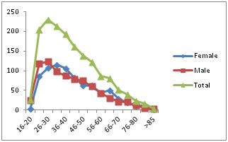
Table 2: Age and sexwise distribution of patients
It was observed that there was male predominance. It was seen that the proportion of fracture was more in male in younger age and the proportion was increasing in female as the age advances.
Figure 1:
Table 3: Distribution of various fractures according to sex
Table 3 (a): Distribution of various fractures according to age
The agewise distribution of fracture showed that fracture of long bones was common in young individuals. Fracture of femur, forearm, humerus and tibia were common in the age group of 21 to 40 years of age. The proportion of fracture femur and humerus were also seen common in elderly individuals also. Table 3(b): Distribution of various fractures according to age
It was seen that fracture of ribs, tarsal, metatarsals pelvis and skull bones were uncommon in the present study.
DISCUSSION The present study was conducted to study the distribution of various fractures observed in the study institution. In the present study total 1590 cases of various fractures were observed in the institute during the study year. It was observed that tibia (29.94%) was the most commonly fractures bone followed by femur (23.08%) and forearm (radius and or ulna) (14.97%). It was observed that there was male predominance. It was seen that the proportion of fracture was more in male in younger age and the proportion was increasing in female as the age advances. Sexwise distribution showed that fracture of femur, forearm and tibia were common among males whereas fracture of hip, spine and humerus were common in females. The agewise distribution of fracture showed that fracture of long bones was common in young individuals. Fracture of femur, forearm, humerus and tibia were common in the age group of 21 to 40 years of age. The proportion of fracture femur and humerus were also seen common in elderly individuals also. B. R. Singeret al10 in their study observed that there was a higher incidence of fractures in men than women in all age groups from 15 to 49 years. The total male incidence is bimodal, with peaks at 20 to 24 years and 90 to 94 years. Females have a smaller peak from 20 to 24 years, with a steady increase later from 46 at 40 to 44 years to 774 per 10 000 population per annum in females aged 90 to 94 years. Liam J Donaldson11 in their study observed that the age specific fracture incidence rates remained below the corresponding male rates until late middle age when the female age groups showed successively higher values than the males. Charles M. et al12 reported that the average age of the patients in their study was 49.1years. The incidence of fractures in men was11.67/1000/year and in women was 10.65/1000/ year. They also demonstrated that a relatively uniform incidence in women up to the menopause and a rapid increase thereafter. However the incidence in men increased in young age and gradually falls until about 60 years of age and again rises again, although the older male peak is lower than the older female peak. It was seen that fracture of ribs, tarsal, metatarsals pelvis and skull bones were uncommon in the present study. T. P. Van Staa13 in their study observed that among women, the radius/ulna (30.2 cases per 10,000 per year) and femur/hip (17.0 per 10,000 per year) were the most frequent fracture sites. In men, the carpal bones (26.2 per 10,000 per year); femur/hip fracture (5.3 per 10,000 per year) were the most common fracture. Varying patterns of fracture incidence were observed with increasing age; whereas some fractures became more common in later life (vertebral, distal forearm, hip, proximal humerus, rib, clavicle, pelvis), others were more frequent in childhood and young adulthood (tibia, fibula, carpus, foot, ankle).
CONCLUSION Thus we conclude that fracture of tibia, femur and forearm (radius and or ulna) were common in adults. The proportion of fracture was more in male in younger age and the proportion was increasing in female as the age advances.
REFERENCES
|
|
 This work is licensed under a Creative Commons Attribution-NonCommercial 4.0 International License.