 Home
HomeOfficial Journals By StatPerson Publication
|
Table of Content - Volume 5 Issue 1 - January 2018
Role of osteotomy and T-Plating [ELLIS] in the treatment of malunited colles fracture
Rajinder Kumar1, Pankaj Miglani2*, A S Sidhu3, Abhishek Garg4, Vishavjeet5
1Associate Professor, 2,4Assistant Professor, 3Professor, 5Sr. Resident, Department of Orthopaedics, Adesh Institute of Medical Sciences and Research, Bathinda, Punjab, INDIA. Email: gargrajinder66@gmail.com, gargrajinder@hotmail.com, dr.pankajmiglani@gmail.com
Abstract Background: Colles Fracture is one of the commonest fracture and malunion is the commonest complication in colles fracture than any other fracture. Since its introduction by Abraham colles in 1814 its management remains a therapeutic challenge. There are various treatment modalities available for treatment of colles fracture with their own merits and demerits. Corrective osteotomy with T Plating is an effective method to treat this malunion, hence this study. Material and Methods: A Study of 25 cases of mal united colles fracture between age group of 19-65 years, treated by corrective osteotomy and volar T-Plate was carried out at AIMSR Bathinda. Movents were advised on very 1st day after surgery and stitches removed on 10-12th post operative day. Follow up was done every 3 weeks till union occurred. The results were evaluated by Fernandez criteria. Results: The average age was 42.12 years, with male predominance and mode of injury was fall on the outstretched hand in majority of patients(92%).Union occurred in 6 weeks in 56% and in 9 weeks in 36% cases. Overall results functionally and cosmetically were excellent to good in 82% of cases. Superficial infection was observed in 8% of cases, stiffness of wrist in 4% and shoulder hand syndrome in 8% of cases, 8% cases had pain at wrist which recovered with physiotherapy but One case had persistent pain and implant needed removal after the union. Conclusion: Conclusion of this study was that by osteotomy and T-Plating with ulna resection(whenever necessary) in malunited colles fracture the pain decreases, movement increases, grip becomes strong and wrist becomes better cosmetically. Key Words: Colles Fracture Distal radius, Malunion, Comminuted Fracture, corrective osteotomy, articular.
The wrist joint is the first link in the complex lever mechanism which connects the hand to the body. Based on the concept that a chain is as strong as its weakest link, It is thus easily seen that the malfunction of the wrist joint impedes the function of one of the two elements which gives mankind its superiority over animals (hand and psyche). Since its description by Abraham colles in 1814, distal radial fracture remains the therapeutic challenge1. Colles fracture is defined as fracture distal end of the radius within 2 cms of the articular surface with dorsal angulation deformity called “dinner park deformity”1,2. This constitutes 17% of all the fractures and 75% of all forearm fractures3. This fracture commonly occurs in elderly patients after the age of 40. The usual mechanism of injury is fall on the outstretched hand. There can be wide variety of displacements of distal end of radius but the common one are impaction, lateral displacement, lateral tilt, dorsal displacement, dorsal tilt and supination. Sometimes it accompanies fracture of the ulnar styloid process which signifies avulsion of the ulnar colletral ligament. AO group has classified these fractures according to their morphological complexity, difficulty in treatment and prognosis. These fractures of the distal end of radius and ulna are classified as (A) Extra-articular fractures (B) Partial-articular fractures (C) Complete-articular fractures. These are further divided to subtypes depending upon the area of involvement and number of fragments. Closed reduction and cast immobilisation has been the mainstay of the treatment of these fractures. But malunion of the fracture and sub luxation or dislocation of the distal radioulnar joint results in poor function and cosmetic results4. Barcon and kurtzke (1953) reviewed over 2000 colles fractures that came before New York state compensation board4. In 7 patients in whom the loss of function was 90% or more and 3% of all the patients had disability of 60% or more. The average permanent loss of function of hand in 2000 cases of colles fracture had been found to be 24%5. Malunion in colles fracture, occurs more common than any other fracture. Loss of palmar tilt of radius in malunion alters the wrists biomechanics, abnormally loading the tenous dorsal ligament complex which results in midcarpal instability with synovitis, pain, weakness and possible articular degeneration. Radial shortening will most likely interfere with function of the distal radioulnar joint and cause pain and limitation of forearm rotation. Malunion may be caused by various factors Like 1.Inaccurate reduction 2. Redisplacement after reduction 3. Comminution of fragments 4.Complete rupture of the distal radioulnar ligament 5. Improper immobilisation 6. Marked crushing of fragments (osteoporosis in elderly). Various methods of treatment available are 1. Ostetomy of the radius and single or dual bone grafting 2. Excision of the lower end of ulna 3. Arthrodesis of the wrist joint 4. Corrective osteotomy and fixation of the lower end of the radius. Darrach (1913) has done partial excision of the lower end of ulna for deformity following colles fracture5. It completely relieved the pain and gave food range of movements, but poor cosmetic results. Campbell described the First biplanar osteotomy and local ulnar bone graft to recreate normal alignment in malunited colles fracture6. The most common forms of surgical fixation are K wires and volar plate fixation. In the last 5 years there has been rapid rise in the use of locking plates which provide improved radiological and/or functional outcomes7-8. The present study was undertaken to evaluate the results of corrective ostetomy and T plating (ELLIS) in the treatment of malunited colles fracture. The present study consisted of 25 cases who were admitted in the Department of Orthopaedics, Adesh Institute of Medical Sciences and Research (AIMSR) Hospital, Bathinda with malunited colle’s fracture with or without other complications. In every case mode of injury, duration and nature of injury, clinical and radiological findings and any previous treatment taken was recorded according to the Performa. The routine investigations in the form of Hb, BT-CT, urine C/E. Blood Urea, S. Creatinine, Blood Sugar, ECG and blood grouping was done. After medical fitness patient was operated under regional/general anaesthesia. Touniquet was applied and limb was cleaned and draped. The lower end of radius was exposed through 7.5 cm long radiovolar incision between the plane of flexor carpi radialis and Palmaris longus. The fibres of pronator quadrates were severed from their origin from the radius and reflected medially .Osteostomy was done at the site of malunion. After correction of the deformity and restoring articular surface,T-shaped Ellis plate was fixed on the volar aspect of the radius. Wound was closed in layers. Patient was advised movement after the 1st day of surgery and check x-ray was taken. Stitches were removed on 10-12th post operative day. Patients were followed up every three weeks till the union had occurred clinically and radiologically. During the follow up, x-ray of the forearm and wrist AP and lateral views was done and patients assessed clinically. The findings were recorded according to the Performa. In repeated follow-up, Physiotherapy and wax bath therapy were given to some patients in physiotherapy department.
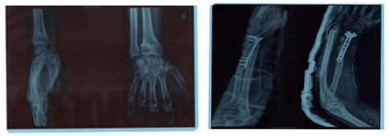
Figure 1: Pre-operative x-ray showing malunited Post-operative x-ray showing corrective Colles fracture osteotomy with excision of lower end of ulna
RESULTS The present study included 25 cases of malunited colles fracture attending the orthopaedics department of AIMSR Bathinda from June 2016 to December 2017.Patients were treated with open reduction and internal fixation with T plate after doing corrective osteomy. 76 % patients fell in the age group of 20-50 years. The oldest was 65 year male and youngest was 19 years old male. The average age was 42.12 years. Male to female ratio was as 60:40 (15 males and 10 females). In 92% (23) cases mode of injury was fall on the outstretched hand. Right side involvement was 76%. In 56 % (14) of cases duration of injury was 9-12 weeks,in 6(24%) was 6-9 weeks and in 5(20%) it was 12-15 weeks. In all the cases there was pain and swelling in the region of wrist extending over the dorsal aspect of hand (Dinner Fork deformity). Tenderness and bony irregularity was found in the lower 1” of radius. Movements of wrist were painful and restricted. 12 patients were given POP cast for 3 weeks after reduction at outside places and others had massages and splints from bone setters. Pre-operative x-rays showed mostly lateral displacement and dorsal tilt of distal fragment. Articular surface was involved in 10 cases (40%) and fracture of the styloid process of ulna was present in 12(48%) cases. Regarding pre-operative complications 24 cases (96%) had pain, 23 (92%) had restricted movements 5(20%) had stiffness of wrist joint, 1 (4%) had carpal tunnel syndrome and 1 (4%) had sudeck,s osteodystophy. 14 (56%) had post operative radiological union at 6 weeks, 9 (36%) at 9 weeks and 2 (8%) had union at 12 weeks. Post operative improvement of motion at wrist i.e. dorsiflexion, palmar flexion, pronation/ supination was 30-35 %. Palmar flexion improvement was deficit mostly(25%),followed by dorsiflexion which had 15% Deficit.Pronation was deficit by 10% and supination by 7.5%. in post-operative complications 4 patients (16%) had pain at wrist, 2 patients (8%) had superficial infection, 1 (4%) had stiffness of wrist and 2(8%) had stiff shoulder(shoulder hand syndrome). The results were evaluated according to the Fernandez criteria11-12.
Table 1: Criteria for evaluation of results
Table 2: Showing clinical results
DISCUSSION Colles fracture is a common fracture especially in the past middle age and most frequent individual fracture of radius. Perfect reduction and maintenance of reduction is essential for obtaining excellent results. If it is not proper or stable, redisplacement of lower fragment can lead to cosmetic deformity, stiffness with functional loss. A malunited colles fracture results in distortion of radial length and the angles of its articular surface in both frontal and saggital planes. Radial shortening with interfere with function of the distal radioulnar joint leading to pain and limitation of rotatory movements of forearm. Decrease in radial inclination will restrict ulnar deviation of hand. Biochemical studies have shows that as the articular surface of the radius tilts dorsally the contact area with scaphoid and lunate also shifts dorsally and decreases in size. This alteration significantly increases the axial load transmitted both to the dorsal aspect of radius and to the triangular fibro cartilage complex,ulnar head area, thus predisposing the wrist to post traumatic arthritis. The priority of any corrective surgery is to reverse the dorsal angle of the articular surface of the radius, normal palmar angle or at least reduce it to right angle to the longitudinal axis of the bone. So keeping in view of the problems faced in malunion of colles fracture, the study of corrective ostotomy and T plate was done in malunited colles fracture. In this study 25 cases of malunited colles fracture were treated in the orthopaedic department of AIMSR Hospital Bathinda. Approval from the research and ethical committe was taken. In the present study age incidence varied from 19-65 years with overall age of 42.12 years. It was most frequent in the age from of 21-50 years i.e. 76%. This may be due to more exposure of younger patients and senile osteoporosis in old patients. The incidence was more common in males 15 (60%) with male to female ratio of 60:40. This may be due to more outdoor activities of males. Commonest mode of injury was fall on the outstretched hand 23 (92%). In 76 % cases Right side was involved. 56 % patients reported with malunion at 9-12 weeks, 24% at 6-9 weeks and 20% at 12-15 weeks. This may be due to rural area, illiteracy and improper guidance by bone setters. Nature of injury was simple in all. No compound fracture was seen. In all 25 cases, there were pain, swelling, tenderness, bony irregularity in lower 1” of radius and dorsum of the hand. Prior treatment with POP cast was taken by 48% and rest 52% had massages and splints from bone settlers. Out of these, most of the patients were uneducated and from rural background which may be a cause of improper treatment. On x-ray 96% has lateral displacement and dorsal tilt and 40 % had interarticular extension. At the time of admission 96% had pain at wrist, 92 % had loss of movement, 32 % had stiffness of wrist and 4% had capal tunnel syndrome with sudeck,s osteodystrophy. In all the 25 cases, Open reduction and internal fixation was done with T Plate fixation after corrective osteotomy. In 2 patients in addition to T-Plating, lower end of ulna was excised to improve the pronation and supination. Follow up was done after 3 weeks and there was radiological union in 56% cases at 6 weeks, 36% in 9 weeks and 8% in 12 weeks. Fernandes (1988) in study of 15 patients had average healing in 6-8 weeks (range 5-8.1 weeks)9-10. Improvement in movement was noted and average palmar flexion increased by 35, dorsiflexion by 33, pronation by 30, and supination increased by 32.The grip strength increased. The deficit of palmar flexion, 68% had reduced to 25%, dorsiflexion 56% to 15%, pronation 47.5 % to 10% and supination 47.5% to 7.5%. This is in comparison with Fernandes (1988) who showed that deficit of pronation 47% and supination 77% had decreased to 12% and 14% respectively9-10. Post-operatively 8% had superficial infection which recovered with appropriate antibiotics and dressings. Stiffness of wrist was in 1 (4%) and shoulder in 2 (8%) of cases. These were having preoperative stiffness but there was improvement with physiotherapy. In one patient, there was persistent pain and plate was removed after 3 months after union and pain subsided with physiotherapy and wax bath therapy. Overall results functionally and cosmetically were excellent to good in 82% of cases, fair in 14% and poor in 4 % cases. Poor results patients had come very late, bones were very ostoportic and was also having sudecks osteodystrophy and was not regular in follow ups. So conclusion of our study was that by osteotomy and T plating with ulnar resection (whenever necessary), the pain decreases, movements and grip improves and wrist looks good cosmetically.
CONCLUSION Corrective osteomy with T plating for malunited colles fracture, though is a difficult procedure through volar approach but gives excellent to good results provided patients come earlier for treatment and at regular follow-ups post operatively.
REFERENCES
|
|
 This work is licensed under a Creative Commons Attribution-NonCommercial 4.0 International License.