 Home
HomeOfficial Journals By StatPerson Publication
|
Table of Content - Volume 8 Issue 1 - October 2018
Outcome evaluation of subtrochanteric femur fractures treated with proximal femoral nails
Soundararajan K
Assistant Professor, Department of Orthopaedics, Vinayaka Mission’s Kirupananda Variyar Medical College and Hospital, Vinayaka Mission’s Research Foundation (Deemed to be University), Salem 636308, Tamil Nadu, INDIA. Email: ksoundar25@gmail.com
Abstract Background: Subtrochanteric femur fractures have demanded special consideration in orthopaedic traumatology, given the high rate of complications associated with their management. The proximal femoral nails were developed as an intramedullary device for the treatment of such fractures. Aim: To evaluate outcome of subtrochanteric femur fractures treated with proximal femoral nails. Material and Methods: A total of 30 Subtrochanteric femur fracture cases were treated with PFNs and followed up for evaluation of functional and radiological outcome. Results: At the end of five months, all except five patients could mobilize independently without any aid. Two patients were using crutch and 3 patients were mobilized using walker. One patient had a superficial infection at the surgical wound site which subsided with parenteral antibiotics. One patient had anaesthesia related complication. We did not come across complications like fracture of femur and failure of fixation. Conclusion: Fractures united in all cases and postoperative functional outcome was satisfactory. PFN could be a preferred implant of choice in treating subtrochanteric fractures especially in elderly since it allows early and stable mobilization. Key Words: Subtrochanteric femur fractures, proximal femoral nails, functional outcome, Postoperative complications.
INTRODUCTION Hip fractures rank in the top ten of all impairments worldwide in terms of loss in disability-adjusted years for people above 50 years old.1Consequences of hip fractures are significant in terms of lives lost and the associated negative impacts on hip fracture patients’ functioning and quality of life. The goal of treatment for hip fractures is to return patients to their pre-fracture level of functions.2 The subtrochanteric fractures are seen usually in 6th and 7th decade, frequently resulting from simple fall. Nowadays due to rapid industrialization and automobiles these fractures are also common in young age group.3 Subtrochanteric femur fractures have demanded special consideration in orthopaedic traumatology, given the high rate of complications associated with their management. As compared to conservative treatment, operative treatment is better tolerated by elderly because of greater comfort, early mobilization of patient, lower morbidity and mortality of patient.4 In subtrochanteric fractures operative treatment is imperative as there is limited role for conservative management.5 The proximal femoral nail (PFN) were developed as an intramedullary device for the treatment of such fractures. In addition to all advantages of an intramedullary nail, it has several other favourable characteristics such as dynamic locking, allows early mobilization, high rotation stability and minimal soft tissue damage.3 The present study was carried out to evaluate outcome of subtrochanteric femur fractures treated with proximal femoral nails.
MATERIAL AND METHODS A total of 30 Subtrochanteric femur fracture cases admitted in Vinayaka Missions Kirupananda Variyar Medical College and Hospital, Vinayaka missions research foundation(deemed to be university) Salem, Tamil Nadu were studied over a period of two years.
Inclusion Criteria
Exclusion Criteria
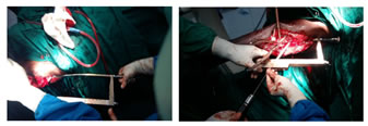
Preoperative Management: The patients were maintained on traction preoperatively in cases whose surgical intervention was delayed for more than two days. All operations were performed under spinal/epidural anaesthesia. High risk patients had thrombosis prophylaxis with low molecular weight heparin subcutaneously during the hospitalization. Transfusion requirements, in-hospital complications, and length of hospital stay were recorded. The patient was positioned supine on the fracture table under spinal or general anesthesia as the condition of the patient permitted. The fracture was reduced by longitudinal traction and the limb was placed in neutral or slight adduction to facilitate nail insertion through the greater trochanter. Prior to positioning and draping, the opposite extremity measurements of rotation and length of this extremity were determined. A straight lateral incision was made from tip of the greater trochanter, extending 4-6 cm proximally; the gluteus maximus muscle was dissected in line with its fibers. Where open reduction was required we extended the incision distally, incising the iliotibial band in line with the skin incision. The entry portal for the PFN was made at the tip of the greater trochanter, halfway between its anterior and posterior extent. A ball tipped guide wire was inserted at the tip of the greater trochanter under C-arm control. The guide wire is advanced into the femoral shaft in such a way that it is located in the middle of the shaft in both directions. In cases where standard PFN was used, we manually reamed the proximal part of the femur with a 14 mm reamer; while where long PFN was used we had to ream the distal femur also with increasing diameters of reamers up to 11 mm. After mounting the appropriate sized nail on the insertion device the nail was introduced manually into the femoral shaft. Via the aiming arm, which was attached to the insertion device, first the guide wire for the neck screw was introduced into the femoral neck in such a way that the 8 mm screw was placed in lower half of the neck on the antero-posterior view and centrally on the lateral view. Thereafter, the guide pin for the 6 mm anti-rotational hip pin was introduced. The hip pin was introduced first with the tip just about 25 mm medial to the fracture line, and then the neck screw of appropriate size was inserted. Afterwards depending on the type of fracture, distal interlocking either statically or dynamically was achieved via the same aiming arm in standard PFN and with free hand in long PFN. The stability of the construct was assessed and wounds were closed in layers over negative suction drain. Antiseptic dressing was done. Per-operatively one dose of antibiotic was also administered. Postoperative Management: Dressing was applied. The limb was elevated in order to reduce swelling and facilitate drainage. Mobilization was started once the patient's condition allows. Weight-bearing was determined by the fracture pattern. In fractures with posteromedial bony contact, graduated weight-bearing were started early. In fractures with high degrees of comminution, weight-bearing was delayed until callus was seen. Figure 1: Mounting of PFN Figure 2: Proximal locking
RESULTS Most number of patients 18 (60%) were in between 30-70 years of age. Seven (23.3%) patients were above 70 years and 5 (16.7%) were between 18-30 years of age. Male patients were more 17 (56.7%) than female patients. Right sided involvement was more 17 (56.7%) when compared to left sided involvement. In our study in 60% of the cases, Road Traffic Accident (RTA) was the cause for the fracture and in remaining 40% cases fall from height was the cause. We did not come across any Type 1 and Type 5 cases and also we came across one patient with Type 4 fracture.
Table 1: Case Distribution according to seinsheimer Classification
Closed reduction was done in 26 (86.7%) of patients and open reduction in 4 (13.3%) of patients. Only two patients needed size 10 Proximal Femoral Nail. The rest had medullary canals appropriate for the size 9 nail. We didn’t use 11 size nail for any of our patients. The operating time for 60% cases was between 2-2.30 hours. The time taken for surgery gradually decreased with familiarity of implant system.
Table 2: Postoperative Independence of Ambulation
At the end of five months, all except five patients could mobilise independently. 2 patients were using crutch and 3 patients were using walker for mobilization. In 9 cases fractures were united between 12-14 weeks, in 21 cases between 14-16 weeks. The average time for radiological union of fracture was between 14 to 16 weeks.
Table 3: Functional Outcome
The type of fracture, according to Seinsheimer classification, did not affect functional outcome. In our study, one patient had superficial wound infection which got subsided with intensive antibiotic therapy and another patient had anaesthetic complication.
Table 4: Complications
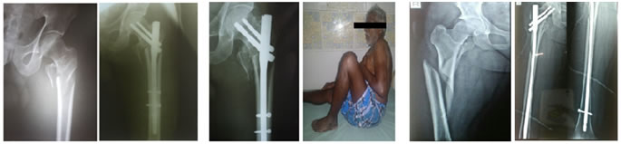
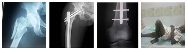
Figure 3 Figure 4 Figure 5 Figure 6 Case 1: Figure 3: Pre-operative Figure 4: Immediate post-operative Figure 5: Two months post-op Case 2: Figure 6 Case 3: Figure 7: DISCUSSION Subtrochanteric fractures of femur are usually the result of high energy trauma. Because of complex stress configuration in this region and its non-homogenous osseous structure and geometry, fractures occur along the path of least resistance through the proximal femur.7 The discussion about the ideal implant for treatment of proximal femoral fractures continues. Closed reduction of the fracture preserves the fracture hematoma, an essential element in the consolidation process. Intra medullary fixation is a more biological fixation and has mechanical benefits over extra medullary fixation.8 The advantages of intramedullary devices over extramedullary ones are less extensive exposure, fewer biomechanical stresses with medial movement of the lever arm and earlier weight bearing.9-12 Intramedullary fixation allows the surgeon to minimize soft tissue dissection thereby reducing surgical trauma, blood loss, infection, and wound complications.13,14 Proximal femoral nail (PFN) has additional anti-rotational screw and the nail tip is specially shaped to reduce the stress and to prevent low energy fracture at the tip of the implant. It greatly reduces the lever arm distance from the reactionary forces generated in the hip joint as a result of movements at the hip joint and increases compressive forces. Some disadvantages of proximal femoral nail which have been reported include cut off of screws in head and neck, lateral migration of proximal screws and femoral medialization.15 In the Christian Boldin study, proximal femoral fractures healed in all 55 patients.16 The longest consolidation time was 5 months which was one month more than the longest time seen in our series. From the mechanical point of view, a combined intramedullary device inserted by means of a minimally invasive procedure seems to be better in elderly patients. All elderly patients did well functionally in our series. We did not come across any implant related complications compared to the 11% seen in the study by Menezes et al.17 In their study, fixation failure occurred in 3 patients(2%), which includes one cut out, one delayed union, one lateral displacement of antirotation screw (total 155 cases).
CONCLUSION In conclusion, PFN is a good implant for subtrochanteric fracture of the femur. The advantages include minimal exposure (closed technique), better stability and early mobilisation. Fractures united in all cases and postoperative functional outcome was satisfactory. PFN could be a preferred implant of choice in treating subtrochanteric fractures especially in elderly since it allows early and stable mobilization.
REFERENCES
|
|
 This work is licensed under a Creative Commons Attribution-NonCommercial 4.0 International License.