Outcome of open fracture of long bones managed with TENS nail
Harshad G Argekar1, Rishabh Jaiswal2*, Santosh N Banshelkikar3, Praveen Jadhav4
1Associate Professor, 2Senior medical officer, 3,4Assistant Professor, Department of Orthopedics, LTMMC Medical college and Sion hospital, Sion, Mumbai, Maharashtra, INDIA.
Email: argekar@gmail.com , drrishabh92@gmail.com
Abstract Background: Open fractures of long bones can involve significant morbidity and are troublesome, as potential for contamination is high in such cases. The correct and timely management of these injuries can benefit patients and lead to more favorable outcomes. Aim: To study the outcome of using TENS Nail as a primary procedure in open trauma long bone. Material and Methods: A total of 30 cases with closed fractures or Grade 1 open fracture where internal fixation could not be done due to poor local and general condition or Grade 2 and 3a open trauma and Grade 3b open trauma cases without gross contamination were managed with TENS nail. Outcome was assessed clinically and radiologically. Results: About 28 (93.4%) patients had radiological alignment while one (3.3%) patient each presented with sagittal plane angulation (>10° procurvatum) and coronal plane angulation (>10° valgus). 4 (13.3%) patients had union on TENS nail. All patients had union after definitive surgery. Conclusion: The better outcome of management suggests that titanium elastic nailing can be a viable option in early management of open fracture long bone with some merit over other procedures.
Key Word: Fracture of long bones, TENS nail, union, complications, outcome
INTRODUCTION
Open fractures of long bones can involve significant morbidity and are troublesome, as the body's protective skin barrier has been broken and the potential for contamination is high. The correct and timely management of these injuries can benefit patients and lead to more favorable outcomes.1 When deciding on the treatment strategy, the treating surgeon must consider the patient's condition, the mechanism of injury, and the fracture type. Goals of open fracture management include the prevention of infection, achievement of bony union, and the restoration of function. Current treatment strategies in the care of open fractures are continuously studied and improved. Plate and screw, external fixator, rigid intra-medullary nails, and TENS (Titanium Elastic nailing system) may be used in surgical treatment. The popularity of TENS has been gradually increasing due to its ease of use and low complication rate. TENS has been used successfully primarily in femur fractures and in various long bone fractures with many published reports in the literature.2-4Hence, the present study was done at our tertiary care centre to study the outcome of using TENS Nail as a primary procedure in open trauma long bone.
MATERIAL AND METHODS
A hospital based prospective study was done with 30 patients with open fracture of long bones over a period of two years.
Sample size
Considering a confidence level of 95% and confidence interval of 18 the number of patients in our study to achieve statistical significance is 30. This was calculated by Survey System (http://www.surveysystem.com/sscalc.htm#one). The Survey System ignores the population size when it is "large" or unknown. Population size is only likely to be a factor when you work with a relatively small and known group of people (e.g., the members of an association). Hence, a sample size of 30 was considered adequate for our study.
Inclusion Criteria
All patients presenting with
- Closed fractures where internal fixation could not be done due to poor local condition, etc.
- Grade 1 open fracture where internal fixation could not be done due to poor local condition, poor general condition, etc.
- Grade 2 and 3a open trauma
- Grade 3b open trauma without gross contamination i.e., cases came to the hospital after 24 hours of trauma and Road Traffic Accident (RTA) cases with crushed bone, bone loss or massive(>1/2 circumference) soft tissue loss
- Age 8-80 years
- Upper and Lower limb long bones fractures
Exclusion criteria
Patients with
- Gross bone loss (more than 1 cm or more than 2/3 of circumference)
- Gross contamination (Grade 3b and 3c)
- Individuals unable to give consent.
- Any comorbidity affecting result outcome
Methodology
As soon as the patient was brought to casualty, patient's airway, breathing and circulation were assessed. Then a complete survey was carried out to rule out other significant injuries. Plain radiographs of AP and lateral views of – the involved extremity including one joint above and one joint below was taken to assess the extent and geometry of fracture. On admission to ward, a detailed history was taken. Routine blood investigations were done for all patients. Patients were operated as early as possible once the general condition of the patient was stable and patient was fit for surgery.
Pre-operative planning of Nail size
Nail length: Lay one of the selected nails were determined that it is of the appropriate length by fluoroscopy. Patients were kept nil by mouth overnight before surgery. Adequate amount of compatible blood was kept ready for any eventuality. The whole of the extremity below the umbilicus, including the genitalia was prepared appropriately. A systemic antibiotic, usually a 3rd generation cephalosporin was administered 1 hour before surgery. Under anaesthesia, closed reduction and internal fixation with TENS nails done under C-arm guidance.
Post-Operative Care And Follow Up
Patients were kept nil orally 4 to 6 hours post operatively IV fluids / blood transfusions were given as needed. Analgesics were given according to the needs of the patient. The limb was kept elevated over a pillow. IV antibiotics were continued for 5 days and switched over to oral antibiotics on the 5th day and continued till the 10th day. Sutures were removed on the 10th postoperative day and patients were discharged. Full weight bearing was started by 8 - 12 weeks depending on the fracture configuration and callus response. Mobilization out of bed without restriction was permitted for patients with isolated injuries. Patients with lower extremity fractures were permitted to bear weight on the upper extremity as tolerated. In case of forearm bone fractures were immobilized the patient for 3wks in a posterior slab followed by allowing ROM exercises for elbow and wrist, sling for another 3more wks. At each follow up patients are assessed clinically, radiologically and the complications were noted.
Statistical Analysis
Appropriate statistical software, including but not restricted to MS Excel, SPSS ver. 20 were used for statistical analysis. Quantitative data was presented with the help of Mean and Standard deviation. Comparison among the study groups was done with the help of unpaired t-test as per-results of normality test. Qualitative data was presented with the help of frequency and percentage table. Association among the study groups was assessed with the help of Fisher test, student ‘t’ test and Chi-Square test. ‘p’ value less than 0.05 was taken as significant.
RESULTS
Majority of the patients (30%) were in the age group of 31-40years followed by 16.7% in the age groups of 21-30 years, 41-50 years and 51-60 years with the mean age of 39.6±14.49 years. There was male preponderance (76.7%) in the study while female patients constituted 23.3% of the study group. There was left sided predominance compared to the right side (56.7% vs. 43.3%).
It was observed that 12 (40%) fractures were Gustilo Grade 2 while 8 (26.7%) and 10(33.3%) fractures were Gustilo Grade 3a and Gustilo Grade 3b. The study included only those Gustilo Grade 3b cases which were not grossly contaminated. The average trauma-surgery interval was 1.8 days. The mean blood loss was 112.5±49.46 ml. The hospital stays of all patients ranged between 8 to 34days. 5 (16.7%) patients were admitted in the hospital for ≤10days while 43.3% and 36.7% patients were admitted in the hospital for 11-20 and 21-30 days respectively. One (3.3%) patient was admitted in the hospital for >30 days. The mean hospital stay was 18.3±7.24 days. The trauma to wound healing duration for 10 (33.3%) patients was ≤2 months, while it was 2-4 months and 4-6 months for 9 (30%) and 11(36.7%) patients respectively. The average trauma–wound healing duration was 3.5±1.36 months. The commencement of mobilisation in 5 (16.7%) patients was 1-3 weeks while it was 3-6 weeks and 6-9 weeks for 21 (70%) and 4 (13.3%) patients respectively. About 28 (93.4%) patients had radiological alignment while 1(3.3%) patient each presented with sagittal plane angulation (>10° procurvatum) and coronal plane angulation (>10° valgus). The primary fixation to wound closure procedure duration for 2 (16.7%) patients was 10-15 days, while it was 16-20 days for 6(50%) and 21-30 days for 4 (33.3%) patients. The average primary fixation to wound closure procedure duration was 18.4±3.82 days. It was observed that no patient required realignment of fixation for wound closure procedure of patient. The removal of primary fixation was done in the same sitting as definitive surgery for all the patients. It was observed that all patients had union after definitive surgery. In our study, 4 (13.3%) patients had union on TENS nail while majority of the patients (86.7%) had no union on TENS nail.
Table 1: Distribution of patients according to Union on TENS Nail
Union of tens nail No. Of patients Percentage
Yes 4 13.3%
No 26 86.7%
Total 30 100%
CASE 1
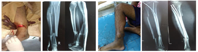
Figure A: Figure B: Figure C: Figure D:
Figure A: Tibia shaft fracture with grade 2 open wound; Figure B: Pre-operative X-rays; Figure C: Post-op a. wound healed after flap surgery; Figure D: Post-op X-rays
CASE 2
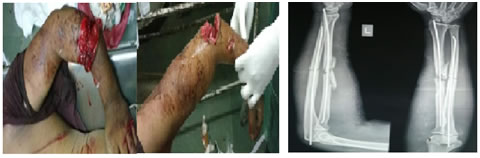
Figure A: Figure B:
Figure A: Grade 3a open fracture associated with assault; Figure B: Preliminary stabilisation using TENS nail
DISCUSSION
In the present study, 12 (40%) fractures were Gustilo Grade 2 while 8 (26.7%) and 10 (33.3%) fractures were Gustilo Grade 3a and Gustilo Grade 3b respectively. In our study, the average trauma-surgery interval was 1.8 days. Wang Q et al5 study on closed reduction and elastic nail fixation for the fibula fracture reported average interval between injury and surgery was 5.8 d (range, 3-22 d).Upadhyay AS et al6 reported nineteen patients were operated within two days of trauma, six patients had a delay in definitive fixation and two patients had head injury and were operated on the 4th and 7th day. The average time interval between admission and surgery was 2.28±1.89 days. Patil et al7 study reported average duration between trauma and surgery was 3.96days. Anjum R et al8 prospective study reported all of the cases were operated within 4 days of injury. The hospital stays of all patients ranged between 8 to 34 days. The mean hospital stay was 18.3±7.24 days. In our study, the commencement of mobilisation in 5 (16.7%) patients was 1-3 weeks while it was 3-6 weeks and 6-9 weeks for 21 (70%) and 4 (13.3%) patients respectively. Wang Q et al5 study reported ankle and knee flexion and extension exercises immediately after surgery, and begun ambulation with crutches and non-weight bearing functional exercise of the affected limb 2 to 3 days after soft tissue swelling disappeared. Patil et al7 study reported 21 (70%) cases were immobilized postoperatively for 6 weeks and such immobilization was for 9 weeks in rest of the 9 (30%) of the cases with an average duration of stay in hospital was 11.6 days. In the present study, 28 (93.4%) patients had radiological alignment while 1(3.3%) patient each presented with sagittal plane angulation (>10° procurvatum) and coronal plane angulation (>10° valgus).Wang Q et al118 reported mean duration of fracture healing in the radiographs was 4.1 months (range, 3-8 months).Upadhyay AS et al6 study reported union rate was 100% in the at the final follow-up. The period of fracture union ranged from 10weeks to 32 weeks, with an average period of 14.98±4.08weeks. There was a single case of delayed union which eventually healed at 32 weeks without any intervention. There were no cases of non-union. In our study, the average primary fixation to wound closure procedure duration was 18.4±3.82 days. It was observed that none of the 12 patients required realignment of fixation for wound closure procedure of patient. The trauma to wound healing duration for 10 (33.3%) patients was ≤2 months, while it was 2-4 months and 4-6 months for 9 (30%) and 11 (36.7%) patients respectively. The average trauma-wound healing duration was 3.5 months. Li SD et al9reported fracture healing time of (12.79±2.52) weeks. The removal of primary fixation for 7 patients was done in the same sitting as definitive surgery for all the patients. The average trauma-definitive surgery duration was 3.7±1.11 months. It was observed that all the 7 patients had union after definitive surgery and none of them had infection after definitive surgery. Wang Q et al5 study reported closed reduction and nail fixation were performed successfully in 18 cases and a limited small incision was made in 5 cases. Primary wound healing was achieved in all patients. No complications such as infection and wound necrosis occurred. Patil et al7 study reported superficial infection in one (3.3%) case. In our study, 4 (13.3%) patients had union on TENS nail while majority of the patients (86.7%) had no union on TENS nail. At the last follow-up visit, the lower-extremity alignment was excellent. Two degrees of varus deformity was found in 3 cases, and 2 degrees of valgus deformity was observed in 2 cases, but there were no serious varus or valgus deformity affecting the lower-extremity function or causing pain. Anjum R et al8 prospective study reported overall complication in 2 (8%) of the cases. The most common complication with titanium nailing is a palpable implant and was present in 8% of the patients. Complications like wound infection, knee stiffness were not encountered. All of the cases returned to full power and range of motion at knee within 16 weeks. Excellent results were observed in 23 (92%) of the patients and good results in 2 (8%) of cases. In a study by Upadhyay AS et al,6 two (10%) patients had nail impingement at the proximal end as it was not buried completely into the bone. There was restriction of shoulder movement (terminal 20 degrees of abduction), and were considered to have moderate functional outcome. One (4%) patient ended up with shoulder stiffness mainly affecting abduction (0-60 degree) and internal rotation (up to lumbosacral junction) at end of 14 months. Upadhyay AS et al6 study showed no incidence of iatrogenic fractures, as titanium has modular of elasticity nearer to the human bone, whereas Enders nail and interlocking nail which are made of stainless steel are stiffer. As a result, titanium nails are easier to negotiate through the bone. As they bend while passing through the bone tension is increased within the nail which improves the three-point fixation. The key distress with respect to titanium elastic nail is its inability to provide suitable rotational stability. The rotational instability can be overcome to an extent with the pre-insertion “C” contouring of nail providing an efficient three- point fixation, distal fanning of nail tips and different entry points for nail insertion. The non-ferromagnetic property of titanium nail further enhances its advantage as it will never interfere with any future MRI (if required). It also reduces the need for a second surgery for implant removal.
CONCLUSION
The use of the TENS nail fixation method in compound fracture long bone has opened up an alternative method of management of these fractures.
Better wound management, patient compliance, decreased hospitalization, low cost of implant, decreased blood loss and operative time with comparable wound healing time suggest that titanium elastic nailing can be a viable option in early management of open fracture long bone with some merit over other procedures.
REFERENCES
1. Court-Brown CM, McQueen MM, Quaba AA. Management of open fractures. St. Louis; London: Mosby; M Dunitz; 1996.
2. Flynn JM, Hresko T, Reynolds RA, Blasier RD, Davidson R, Kasser J. Titanium elastic nails for pediatric femur fractures a multicenter study of early results with analysis of complications. J PediatrOrthop 2001; 21:4-8.
3. Luhmann S, Schootman M, Schoenecker Pl, Dobbs MB, Gordon JE. Complications of titanium elastic nails for pediatric femoral shaft fractures. J PediatrOrthop2003; 23: 443-447.
4. Sink E, Gralla J, Repine M. Complications of paediatric femur fractures treated with titanium elastic nails: a comparison of fracture types. J PediatrOrthop 2005; 25: 577-580.
5. Wang Q, Hong-Guang X, Yin-Chang Z, and Li-Jun D. Elastic nails for fibular fracture in adult tibiofibular fractures. Int J ClinExp Med. 2015; 8(6).
6. Upadhyay AS, Lil NA. Use of Titanium Elastic Nails in the Adult DiaphysealHumerus Fractures. Malaysian Orthopaedic Journal 2017; 7(2).
7. Patil S et al. Prospective Study in treatment of long bone fracture in children by Titanium Elastic Nailing stabilization. Int J Biol Med Res. 2014; 5(4): 4479-4490.
8. Anjum R, Raina P, Singh S, Hackla S, Malhotra S. Fixation of Paediatrictibial fractures with TENS; A prospective study. IntJ Advanced Research. 2015; 3(5):251-254.
9. Li SD, Xu C, Tong PJ. Application of external fixator combined with damage control treatment for open fracture of the extremities. Zhongguogu Shang. China JOrthopTraumatol 2015, 28(2):130-135.
 Home
Home This work is licensed under a Creative Commons Attribution-NonCommercial 4.0 International License.