 Home
HomeOfficial Journals By StatPerson Publication
|
Table of Content - Volume 8 Issue 2 - November 2018
A study of clinicoradiological outcome of distal tibia fractures treated with various modalities
Parag M Tank1*, Rajesh V Chawda2, Nimish B Patel3, Vijay J Patel4, Chirag P Parmar5*
1Assistant Professor, 2Associate Professor, 3Professor, 4,5PG Resident, Department of Orthopaedics, N H L Medical College, S C L Hospital, Saraspur, Ahmedabad, Gujrat, INDIA. Email: chiragparmar51292@gmail.com
Abstract Background: Distal tibia fractures are seen as very high energy trauma, in the same way it is curiously enough to understand and outlook the in-depth management. Quite evidently it is worthwhile to conclude the exact geometry of the injury likewise its rational treatment protocol. Soft tissue around ankle is of utmost importance if neglected may lead to serious problems. Method Prospective study of 13 patients of AO type A 12 and type C 1 patient distal tibia fractures between ages 25-70 years sustained injury following road accidents. 2 patients had Gustilo Anderson type I while 2 had type II wounds. We did initial damage control ankle spanning external fixator adjunct to fibula nail followed by definitive LCP in 8 patients, IM nail in 3 and augmented external fixator in 2 patients. Follow up of the patients at 12-18 months. Result Mean radiological union was achieved at 13 weeks. AOFAS score was excellent in 7 cases while good in 4 patients. 1 patient of open grade II wound had superficial infection.1 patient had pain while walking. 1 patient had implant impingement. A patient treated with IM nail had delayed union. No secondary bone graft surgery, soft tissue flaps needed. No rotational malalignment observed. Conclusion In the ultimate analysis of fracture classification and soft tissue trauma it is wise to do staged surgery in high degree tissue and bony insult in these injuries. IM nail, LCP and definitive external fixator are mainstay of resort. Key Word: distal tibia, LCP, IM nailing, AOFAS.
INTRODUCTION Carrying approximately 10% of skeletal trauma, distal tibia fractures are amenable to very high expertise to treat and manage. The rescue to these axial and rotational loading induced injuries is to respect and handling the soft tissue around ankle. Victorious outcome shall only be achieved following staged minimum open procedures and early rehabilitation. Consensus has been long to address whether to operate or not and if so then what kind of implants are there for remedy. Intramedullary nails are now utilised in these injuries in selected cases. But problems remain the same with rotational and angular malalignment. Tibial metaphyseal plates are mainstay following AO locked plates armamentarium which necessitate expertise and proper tissue handling while inserting. Precarious blood supply and soft tissue and muscle around distal tibia make it vulnerable to heal after injury. Additional surgical tissue handling further jeopardise the venous and neural network. Comorbidities like diabetes, hypertension, varicosities around ankle, hyper cholesterolaemia and smoking habit still delay healing. Compartment syndrome, delayed presentation to emergency set up and excessive swelling hamper the definitive procedures not only late but also functional outcome compromised. Careful dissection of distal tibia while anteromedial or anterolateral approach and switch to mippo procedures is of gold standard. External fixator has advantages over open techniques related to tissue handling but has cons to substandard reduction, pin tract infections and ankle stiffness. Intramedullary nailing with latest designs promise to stabilise the most distal small fragment in some fracture geometry. Still improvements and innovations are required to understand and execute the technical aspect of management of these injuries.
MATERIAL AND METHOD Our study had 13 patients of distal tibia fractures sustained between June 2015 to January 2017. We include 9 male patients and 4 females which met with injury which was classified according to AO OTA distal tibia classification system. All patients were undergone surgery by a group of surgeons after preoperative thorough radiological skeletal survey following these high energy trauma. Demographic assessment were done considering age, sex, mode of injury.(TABLE 1). No CT scans were done for any of the patients of these injury. Patients were counselled for the need of staged surgery if severe swelling, open injuries and comorbidities. All patients were operated under spinal anaesthesia after informed written surgical consent and accounted after institutional ethical committee review enrolment. Inclusion criteria
Exclusion criteria
After full assessment of the radiology and local skin consistency we planned distal tibia anatomical locking plate. Where skin condition didn’t favour for direct open reduction we switched to distractor two steinmann pin ankle spanning fixator. 3 patients were undergone distal tibia tip interlocking ntramedullary nail following primary lateral column fibula rush pin fixation and external fixator for few days. Two patients were treated with temporary ankle spanning fixator followed by two plane definitive fixator. Skin incisions taken at anteromedial aspect of distal tibia starting from tip of medial malleolus to crest of lower tibia. Saphenous neurovascular bundle identified, tagged and retracted. Usually fracture site was opened and reduced under direct vision. Multifragmentary fracture lines were not addressed each as soft tissues of this jeopardised segment could be at stake. But two to three posteromedial and posterior fragments were held with interfragmentary 4.5mm cortical screws. As lateral columns were fixed initially in emergency setting along with fixator, the final reduction manoeuvres were significantly reduced. Anatomical distal tibia low profile locking compression plates were slid from below. Initially plate was fixed with proximal screw and distally with Kirschner wires. Having confirmed the alignment, length, anatomical geometry of fracture and adequate plate placement under image intensifier, final locking head screws were fixed. Rotational mal-alignment were checked each time with measuring and visualising plum line of the limb and true limb axis comparing both sides intra-operatively. After checking final image, wash was given to the wound and closure done in layers with 2-0 absorbable suture and 3-0 non-absorbable skin suture. Compression bandage applied and limb elevated in below knee splint support. In the outset of the patients of type A AO classification, we did secondary farthest tip locking intramedullary interlocking nails. Routine intramedullary nail entry selected at parapatellar region and nail inserted of adequate size diameter and length after fully reaming of the medullary canal with flexible reamers just above the ankle plafond. Again rotation of the limb was the paramount importance as we advanced the nail each task of propagation was checked with plum line of the limb. No varus valgus malalignment was encountered as lateral column were already fixed in the golden period per se. no heavy traction table attachments were required in any of the patients. We did definitive external fixator for two patients as had gustillo anderson type 2 open injuries. Fracture alignment was achieved with temporary spanning fixator and biplanar pins were fixed through the universal clamps and frame remained after achieving alignment and rotation. Wound was debrided and skin grafted with split graft. Dressing was applied and limb splinted with below knee plaster slab. Patients were regularly dressed on day 2, day 7 and day 15. Sutures were removed at post operative day 15. Xrays were taken on day 2, after a month and after 3 and 6 months. Quadriceps and hamstrings exercises were started immediately from day one. Definitive fixators were removed a month after appearance of radiological union. Non contact walking was started on day two if general condition permit. Partial weight bearing was started after 10-12 weeks and full weight walking was started after 14-18 weeks postoperatively. Evaluation of the questionnaires regarding improvement and result were started at 4-6 weeks and at 6 months and lastly at a year post surgery. American foot and ankle scores were measured and written each time visit but three visits as above were taken as a final.
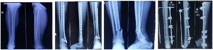
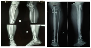
Figure 1 Figure 2 Figure 3 Figure 4 Figure 5 Figure 6 Legend Figure 1: m/50 patient xray AP and lateral view shows distal tibia fibula fracture type A3; Figure 2: Same patient treated with MIPPO and fibula rush pin;Figure 3: 25/M xray shows anterior and Posterior view of distal tibia type A3andOG-2; Figure 4: Same patient treated with external Fixator+rush pin; Figure 5: 27/M xray shows anterior and posterior view dista; tibia fibula fracture type A3; Figure 6: same patient treated with tibia interlock intra-medullary nail and fibula rush pin.
RESULTS Study was conducted at our institution between June 2015 to January 2017 with average follow up 14 months(12-18 months). We did distal tibia fracture study in 13 patients among them 9 patients(69.2%) were males and 4 patients (30.7%) were females. 8 patients(61.5%) had injury in right leg while 5 patients(38.4%) had left leg injury. Average age of the study patients was 44 years(range 25-70 years). 10 patients (76.9%) had injury of their leg after met with a road traffic accident. 1 patient(7.6%) had house fall and 2 patients(15.3%) had injured his leg following fall from height. 2 patients(15.3%) had Gustilio Andersion open type I injury while 2 patients(15.3%) had type II open injury. 7 patients(53.8%) had associated lower shaft fibula fractures. 1 patient(7.6%) had L1 compression injury without neuro deficit. 12 patients(92.3%) had AO OTA type A1, A2 and A3 while 1 patient had type C2 pattern of injury. 3 patients (23%) had been undergone distractor ankle spanning two steinmann pin fixator along with percutaneous rush pin fixation for lower third fibula fractures in emergency room. Definitive surgery was undertaken at an average 4 days (range 3-10 days)(FIGURE 3 and 4). 2 patients(15.3%) had definitive external fixator till healing and weight bearing rehabilitation which was removed at the end of 16-18 weeks. 3 patients(23.7%) had farthest tip locking intramedullary interlocking nail with rush pin in fibula fracture.(FIGURE 5,6) We observed the patients for skin wounds and swelling for 3 weeks. Pin tract dressings and rehabilitation physiotherapy programme continued until removal of fixator and full weight bearing mobilisation.We observed radiological union at mean 13 weeks (range 10-18 weeks). Union of multifragmentary fractures showed longer healing. American ankle and foot scores were observed at 4-6weeks, 6 months and at a year considering the pain, rom and ankle and complications related to surgery as well as more comminuted pattern of metaphyseal fracture. Excellent result was observed in 7 cases (53.8%), good result in 4 cases (30.7%) while fair result was seen in 2 patients(15.3%).(TABLE 2) We observed pain at terminal ankle dorsiflexion and at full weight bearing mobilisation which required stick support for 6 months in 1 patient(7.6%). 1 patient with LCP had pain while stress test at ankle which didn’t show tibiofibular distraction on mortise view. Arthritis at posterior tibiotalar joint was observed in 2 patient of AO type C2 fracture pattern at final follow up. 1 patient with LCP had implant impingement as patient was thin in built. 1 patient with open grade II injury treated with definitive external fixator had superficial infection which resolved with regular dressing and broad spectrum antibiotics for 8 weeks. 1 patient with type A3 AO fracture treated with tip locking interlock nail had delayed union on 6 months follow up which showed consolidation at 14- 18 months. No bone graft was needed as a second procedure in any of the cases. Rotational and angular malalignment, limb length discrepancy were not observed in any of our cases.
Table 1: (Demographic Table)
M- MALE, F-FEMALE, RTA-road traffic accident, DTLP-distal tibia locking plate, I.M-intramedullary, MIPPO-minimal invasive percutaneous plate osteosynthesis. Table 2:
DISCUSSION Distal tibia fractures management has been evolved drastically after accepting the fact of sparse blood supply at and around ankle which make the fracture as well as overlying skin non healing or delayed healing. Open reduction and internal fixation of metaphyseal distal tibia fractures have been suggested by many surgeons since ages. Newer generation and AO design nails and choice of locking in distal part of the nail beyond the fracture ends confer implant of recommendation in selected cases. Since open injuries at ankle require temporary damage frame immobilisation and skin coverage with split grafting or flap render a surgeon contemplating whether switch to internal implant or definitive external fixator. Intramedullary nailing in distal tibia fractures has some less intrinsic stability, decreased load sharing, less stability of construct lead to multiple point kinking of distal screws.1 we had one case of delayed union in our study treated with fibula rush pin and intramedullary nailing. This happened owing to more comminuted metaphyseal fracture and segmental blood supply to each fragments. No distraction was kept at the time of surgery but accepted compression which is achieved at mid shaft was not seen due to more comminution. Cantilever bending is much more achieved in nail fixation rather than medial plate. Locked plate construct along with lateral column fixation as fibula fracture stabilization demonstrated excellent torsional stiffness and no displacement of fracture as well as fixed angle stability with screws near to the fracture compared to nail where screws are far.(FIGURE 1,2) But intramedullary nail fixation along with fibula plate or nail has robust length, stiffness, stability and decreased valgus malunion as well.2 Gulabi et al3 had described 2 superficial infections in open platinng group(9.1%) which was recovered with local wound dressings. Infections are generally associated with these fractures owing to precarious blood supply to the lower segment of tibia, high velocity injury which further hamper circulation, swelling, and delayed presentation at the emergency care centre. Madhuchandra et al4 study has suggested plating in distal tibia type C AO is better for anatomical reduction and good functional outcome nevertheless nailing is as good in type A and selected type B cases. Vaza et al5 had conducted study in 40 cases of distal tibia fractures between age group of 18-65 years with majority of road traffic accidents as mode of injury. 90 % of cases had associated fibula fractures. Average time of union was 23 weeks in group 1 plating and 26 weeks in group 2 nailing. Gupta et al6 involved 79 patients of distal tibia fractures treated with distal tibial metaphyseal as well as medial tibial LCP had two cases of delayed union. All fractures were treated with fibula fixation first. Five patients had secondary bone grafting. Results showed better implant construct with respect to healing of bone and soft tissue and functional outcomes. We had 8 patients with metaphyseal anatomical locking compression plate with no soft tissue complications except delayed suture removal in some cases. Concha Sandoval JM et al7 divided their study patients in three groups according to AO type A, B and C. He demonstrated consolidation time ranges from 14 to 17.4 weeks. He concluded plate osteosynthesis is better option for acquiring correction of fracture geometry and soft tissue healing. We did one third tubular plate in C2 fracture and 4 mm cancellous cannulated screw in lower end fibula fracture which showed ankle arthrosis at final follow up. Francois et al8 retrospective study of 10 patients treated with percutaneous plating for ankle plafond showed no significant soft tissue problems, two cases with delayed union and all fracture healed within a year. Patients were explained before and even after surgery about need of bone grafting if any situation arose of delayed and non-union. We didn’t find any data regarding primary bone grafting. Still a matter of consensus at what point of time these high velocity fractures behave particularly at consolidation and healing of multifragmentary geometry because at every follow up after weight bearing it showed union related lag at radiology. Ravindra et al9 emphasised LCP a safe and effective line of management for AO type A, B and C fractures related to union and few complications. Rabari et al10 had prospective comparative study of 73 patients between intramedullary nailing and plating for distal extra articular distal tibia fractures with mean age of the patients was 39.9 years. It suggested AOFAS score 58.9% in plating and 41.1% in nailing group and recommended plating has superior outcomes. Goel et al11 treated 18 patients of AO type A fractures with ankle spanning external fixator and fibula fixation with limited internal fixation like kirschner wire and lag screw had average healing time 16-18 weeks with 83.3% good to excellent results without any major complications. Cui et al12 conducted a meta-analysis between two stage ORIF and limited internal fixation external fixator and concluded no major difference in union and infections. Egol et al13 instituted intermediate placement of spanning external fixator on the day of admission in soft tissue compromised patients of these high energy injuries in intra articular and metadiaphyseal fractures with mean age was 47 years with final WOMAC score 91. Early osseous stabilisation with spanning external fixator create a good skin environment and fracture alignment with length which is mandatory and foundation for definitive surgery or persistence of same fixator as final construct. Sirkin et al14 studied 226 pilon fractures with group 1 close injuries group 2 open injuries had treatment protocol of fibula open reduction and internal fixation and spanning external fixator for pilon fractures. Following soft tissue oedema subsidence, definitive plate fixation attempted. There were three soft tissue infection noted on follow up which were recovered with serial debridement. Soft tissue swelling and tissue insult should be quiet when external fixator applied initially to achieve fibular length, articular alignment. Khuntia et al15 also stressed the importance of fibular fixation to achieve length of the limb and biomechanical advantage of minimal weight transmission. Some literatures have also entioned delayed tibial healing in subset of fibular rigid fixation group owing to impede cyclical compression. Our technique of patient selection and initial damage control frame which span the ankle significantly reduce the skin swelling, restoration of fibular length adjunct with rush pin fixation, articular alignment in type C injuries. Secondary whatever the procedures performed by clinician, it shift the paradigm to lesser complications especially infection, wound dehiscence and late valgus deformity. Interosseous membrane which anchors tibia with fibula, also regain its resilience after fixation of fibula and external fixator on the same day of injury. Furthermore the locking plate or nail fixation as definitive stage become easy and technically sound as necessity of opening the fracture become meagre. onetheless, patient has to undergo two stage surgery and bear cost of two inventory, the functional final outcome improve the quality of life and abolish the rate of complications of late loss of reduction and malalignment.
CONCLUSION In spite of the fact that direct open reduction has more advantage in fracture management, nevertheless, distal tibia should be tackle with minimum soft tissue disruption with MIPPO plate, intramedullary evices or external fixators. Above and beyond the cost and time of hospital stay it is also not irrational to stage the distal tibia fracture management protocol. With due respect to lateral column fixation initially confer ease of secondary procedure with pliable skin around ankle which exhibit good to excellent outcome.
REFERENCES
|
|
 This work is licensed under a Creative Commons Attribution-NonCommercial 4.0 International License.