 Home
HomeOfficial Journals By StatPerson Publication
|
Table of Content - Volume 8 Issue 2 - November 2018
Clinical profile of patients admitted with supracondylar fracture of femur: A study from Swasthiyog Pratishthan, Miraj
Kiran P Paknikar1*, Shekhar Malve2, G S Kulkarni3, M G Kulkarni4, S G Kilkarni5, Anant A Takalkar6
1Consultant Orthopedic and Spine Surgeon, Ankur Hospital, Vasai, Maharashtra, INDIA. 2Lecturer, 3Consultant Orthopedic Surgeon, 4,5Professor, Department of Orthopedics, Post graduate Institute of Swasthiyog Pratishthan, Miraj, Maharashtra, INDIA. 6Professor, Department of Community Medicine, MIMSR Medical College, Latur, Maharashtra, INDIA. Email: paknikar.kiran1@gmail.com
Abstract Background: Supracondylar and intercondylar fractures of femur are very often difficult to treat and they are notorious for many complications. In the supra and intercondylar fractures of femur particularly with intra articular extension, patient may develop stiffness of knee, shorting, rotational deformities, internal derangement of knee with instability, varus and valgus deformities which affect patient’s routine life style. Objectives: To study the clinical profile of cases admitted with supracondylar fracture of femur and its management. Methodology: Prospective Longitudinal observational study Conducted at Post Graduate Institute of Swasthiyog Pratishthan, Miraj, Maharashtra involving 50 patients with supracondylar fracture. The fractures were classified as a supracondylar femur fracture (AO/OTA type 33) (A-C). Fractures that were supracondylar with significant proximal fracture extension were classified as an AO/OTA type 33 fracture unless there was a separate diaphyseal fracture. Results: Majority of patients were from 30-39 years age group i.e. 32%. Majority of patients were males i.e. 46 (92)%. In majority of cases, road traffic injury is the commonly observed cause of fracture i.e. in 47 (94%). 8(16%) patients had type A fractures. 1 (2%) patients had type B fractures and 41(82%) patients had type C fractures. Majority of them i.e. 52% cases had ROM between 70-100 degrees. Conclusion: Supracondylar fracture seen in middle age group with male preponderance. Common cause is RTA. LCP provides stable construct especially in cases with metaphyseal comminution and enables early mobilization. Key Word: supracondylar fracture, femur
INTRODUCTION Over the centuries from ancient ages to the present age of advanced technology there have been many changes in the life style of mankind. Industrialization and the fast pace of life have brought both comforts and catastrophic road traffic accidents, crippling many young lives. Supracondylar and intercondylar fractures of femur are very often difficult to treat and they are notorious for many complications. In the supra and intercondylar fractures of femur particularly with intra articular extension, patient may develop stiffness of knee, shorting, rotational deformities, internal derangement of knee with instability, varus and valgus deformities which affect patient’s routine life style. If such fractures are not properly treated the individual becomes crippled, thus affecting the country’s working class and nations economy.1,2 Surgical treatment of supracondylar or intercondylar distal femoral fractures (AO/OTA types 33-A To 33-C) remains a significant surgical challenge with significant complication rates.1,7 Adverse events include infection, decreased range of motion, need for bone grafting, malunion, and nonunion.1,2,4,7 Emphasis on preservation of osseous vascularity utilizing indirect reduction techniques has led to increased union rates without bone grafting.8, 9
METHODOLOGY Study setting: Post Graduate Institute of Swasthiyog Pratishthan, Miraj, Maharashtra. Study design: Prospective Longitudinal observational study. Sample size: 50 patients with supracondylar fracture. Study period: August 2007 to September 2009. Sampling method: All patients with confirmed diagnosis of supracondylar fracture reported to our institution during above-mentioned period. Selection criteria: This study involved 50 patients who sustained fractures of AO/OTA type 33(A to C) both closed and compound according to Gustilo and Anderson classification were chosen. Age group ranging from 20-80 years who were treated using (LCP) locking compression plate in post graduate institute of Swasthiyog Pratishthan Miraj operated between August 2007 to September 2009. All fractures were classified according to the AO/OTA classification system. Fracture demographics: The fractures were classified as a supracondylar femur fracture (AO/OTA type 33) (A-C). Fractures that were supracondylar with significant proximal fracture extension were classified as an AO/OTA type 33 fracture unless there was a separate diaphyseal fracture.
RESULTS Table 1: Distribution of study population according to age
In our study majority of patients with supracondylar fracture were from 30-39 years age group i.e. 32% and 24% from 20-29 years as well as 40-49 years age group each
Majority of patients were males i.e. 46 (92)% and remaining were females i.e. 8% In our study we found preponderance of male population with male to female ratio as 11.5:1. 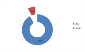 Table 2: Distribution of subjects according to type of injury
Closed type of injury was seen in 27 patients i.e. 54% whereas in 23 patients i.e. 46% it was compound in type.
Table 3: Distribution of subjects according to mode of injury
In majority of cases, road traffic injury is the commonly observed cause of fracture i.e. in 47 (94%). 6% patients reported fall at home. Table 4: Distribution according to fracture subtype
The cases were classified according to AO/OTA classification. 8(16%) patients had type A fractures. 1 (2%) patients had type B fractures and 41(82%) patients had type C fractures
Table 5: Distribution according to postoperative range of motion (ROM)
Out of the total number of cases, majority of them i.e. 52% cases had ROM between 70-100 degree followed by 28% between 0-70 degree. 20% had more than 100 degrees.
DISCUSSION A supracondylar fracture of the femur is a grave injury that for years represented an unsolved problem in trauma and was considered to result almost always in varying degrees of permanent disability. It was felt that the fate of the joint was determined by the injury rather than by its treatment. Closed procedures were almost always used in treatment, and consisted principally of splinting and traction. Open reduction and internal fixation were attempted from time to time, but the results were largely unsatisfactory, because the techniques of internal fixation and the devices available did not allow stable fixation, which would allow early motion without deformity and nonunion. In our study majority of patients with supracondylar fracture were from 30-39 years age group i.e. 32% and 24% from 20-29 years as well as 40-49 years age group each. Majority of patients were males i.e. 46 (92) % and remaining were females i.e. 8%. In our study we found preponderance of male population with male to female ratio as 11.5:1 Ramana SSV et al10 studied involving 18 males and 2 females age ranging from 20 years to 65 years with an average age 40 years. Average age for males is 28.9 years and average agr for females is 25 years. In our study, closed type of injury was seen in 27 patients i.e. 54% whereas in 23 patients i.e. 46% it was compound in type. In majority of cases, road traffic injury is the commonly observed cause of fracture i.e. in 47 (94%). 6% patients reported fall at home. The cases were classified according to AO/OTA classification. 8(16%) patients had type A fractures. 1(2%) patients had type B fractures and 41(82%) patients had type C fractures Ramana SSV et al10 included 17 fractures that were due to road traffic accidents and 3 cases due to fall from varying heights. Among 20 cases there are 3 compound fractures 15% and in them 2 cases were type 1 66% 1 case was type 2 33%.for classification of open fractures we have used gustilo Anderson classification. He included muller’s type A and C fractures. Sub groups are type A 1-3 cases, A2-2 cases, type A 3-5 cases, type C 1-4 cases, type C 2-3 cases and type C3-1 cases. In our study, the mode of treatment was different for each type. Type A1 were treated by compression system, type A2 and A3 were treated by splinting system. Type B fractures were treated by compression system. Type C fractures were treated by anatomic reconstruction of articular surface and splinting system.2A1 were treated by compression system.2A2 and 4A3 were treated by splinting system. 1B3 was treated by compression system. 3C1, 24C2, 14 C3, were treated by reconstruction of articular surface and splinting system. In our study we found that out of the total number of cases, majority of them i.e. 52% cases had range of movement between 70-100 degree followed by 28% between 0-70 degree. 20% had more than 100 degrees. Ramana SSV et al10 in his series of present study of 20 fractures, 12 cases were treated with dynamic condylar screws,4 cases were treated with retrograde intramedullary nailing and in 4 cases with locking compression plate. Average ROM in their study was 74.28 degrees and type A fractures 98.46 degrees and type C fractures 71.41 degrees.
CONCLUSION Supracondylar fracture seen in middle age group with male preponderance Common cause is RTA. Locking compression plate is an ideal implant for fixation of supracondylar fracture of femur 33 (A-C) especially in C3 type where articular comminution is present. LCP provides stable construct especially in cases with metaphyseal comminution and enables early mobilization.
REFERENCES
|
|
|||||||||||||||||||||||||||||||||||||||||||||||||||||||||||||||||||||||||||||||||||||||||||||||||
 This work is licensed under a Creative Commons Attribution-NonCommercial 4.0 International License.