 Home
HomeOfficial Journals By StatPerson Publication
|
Table of Content - Volume 8 Issue 3 - December 2018
Clinical and functional outcome of recession of gastrosoleus by Vulpius procedure in spastic diplegia type cerebral palsy children
Bharati Pankaj DeokarSharma1, Pankaj Nandkishor Sharma2*
1Assistant Professor, 2Senior Resident, Department of Orthopaedics, B.K.L.Walawalkar Rural Medical College and Hospital, Kasarwadi, Dervan. 415606 INDIA. Email: bharati.deokar@gmail.com
Abstract Background: Equinus deformity is the most common foot deformity in patients with cerebral palsy, affecting 70% of children. There are many different procedures to correct the equinus deformity with most having insufficient research to support. Aim: To evaluate clinical and functional outcome of gastrosoleus recession by Vulpius technique to correct equinus deformity in spastic diplegia type cerebral palsy patients. Material and Methods: A total of 35 spastic diplegia type cerebral palsy patients with equinus deformity were treated with recession of gastrosoleus at its musculotendinous junction by Vulpius method. The patients were classified by Physician Rating Scale, Modified Ashworth Scale, Gross Motor Function Classification System, Expanded and Revised (GMFCS – E and R) and Modified Functional Walking Scale. Results: In mid-stance all patients improved to heel-toe or flat-foot except one. No change was observed in GMFCS-ER grades post-operatively. All the patients operated by our technique had plantigrade foot except one at end of one-year follow-up. All patients had significant improvement as all moved to next grade respectively. In all patients, spasticity decreased by at least one grade. Out of 34 patients, 26 required external support pre-operatively while only 11 required it post operatively at end of one-year. Conclusion: Gastrosoleus recession by Vulpius method is an effective procedure in treatment of spastic diplegia type cerebral palsy patients with equinus. Patient had good improvement with minimal complications. Key Word: Cerebral palsy, spastic diplegia, equinus, gastrosoleus recession, Vulpius method
INTRODUCTION In cerebral palsy, the muscles of children are smaller compared with unaffected children. Growth of the muscle tendon units lags behind growth of long bones, resulting in deforming contractures.1,2Spastic is the most common form of cerebral palsy, constituting approximately 80% of cases, and usually is associated with injury to the pyramidal tracts in the immature brain. Diplegia is the most common anatomical type of cerebral palsy, constituting approximately 50% of all cases. Patients with diplegia have motor abnormalities in all four extremities, with the lower extremities more affected than the upper. This type of cerebral palsy is most common in premature infants; intelligence usually being normal.3 In spastic diplegia, when child learns to walk he most commonly develops a ‘Jump gait’ in which the child walks with hips in flexion, knees in flexion and ankles in plantar flexion as if ready to jump.4Foot deformities caused by altered or abnormal muscle forces are common in patients with cerebral palsy, with approximately 70% to 90% of children affected.3 Equinus deformity is the most common foot deformity in patients with cerebral palsy, affecting 70% of children.3It is defined as the inability to dorsiflex the foot above plantigrade, with the hindfoot in neutral and knee extended.5 Surgical treatment is a rapid and dramatic means of altering the structure and functioning of the musculoskeletal system in children with cerebral palsy. It is almost always the only effective means of treatment when fixed myostatic contracture exists. Lengthening a muscle also weakens it. This may help with muscle balance around a joint. There are many different procedures to correct the equinus deformity with most having insufficient research to support. There is inadequate evidence to state a clear preference for any single surgical procedure. The studies evaluating the correction of equinus deformity by gastrocnemius recession using the Vulpius method have been few even though the method has been described a century back. In our study, we carried out gastrosoleus recession by Vulpius technique to correct equinus deformity in spastic diplegia type cerebral palsy patients and evaluated its clinical and functional outcome.
MATERIAL AND METHODS This prospective study was carried out at SASMCT'S Post Graduate Institute of Orthopaedics and Trauma, Sangli, Maharashtra, India over a period of two years. During the study period all patients of spastic diplegia type cerebral palsy planned for gastrosoleus recession procedure were screened with the inclusion and exclusion criteria. Informed, written consent was taken for all patients that fit the inclusion criteria and all patients willing for assessment were included. The protocol was reviewed and approved by an Institutional Ethics Committee. Inclusion Criteria
Exclusion Criteria
History of all included patients was taken with regard to birth weight, gestational age at birth and any complications that occurred after birth and documentation of motor milestones were made. Physical examination: Specific items were included in the physical examination of a child with cerebral palsy. Tests were done for diagnosing tightness of muscles such asIliopsoas, Adductors, Gracillis, Hamstring, Medial hamstring and Tendoachilles. Thorough clinical examination of all patients was done and the treatment was instituted according to the need of patient. Appropriate physiotherapy was given to early detected cases. Silverskiold test was performed on each patient but irrespective of its result all patients having equinus and qualifying the inclusion criteria were operated. Pre-operative routine laboratory investigations were done. All patients were identified and classified by Physician Rating Scale, Modified Modified Ashworth Scale, Gross Motor Function Classification System, Expanded and Revised(GMFCS – E and R)and Modified Functional Walking Scale. Surgical technique: Recession of gastrosoleus at its musculotendinous junction by Vulpius method. Post operatively, patients were given above knee cast for one month. They were advised full weight bearing walking from day 2 post-operative. After one month, cast was removed and aggressive physiotherapy for lower limb movements was given. Follow up: The patients included in the study were followed up at regular intervals at 4 weeks, 3 months, 6 months and 1 year. At each follow-up clinical examination for evaluation for maximum passive Ankle dorsiflexion, physician rating scale assessment, GMFCS-ER Classification, Modified Ashworth scale, Modified functional walking scale, Brace requirement, Support requirement for mobility and Parent satisfaction.
RESULTS A total of 35 patients completed the follow up whereas one patient was lost to follow-up hence, he was discarded while analyzing the data. Patients were selected from age group of 5 years and above upto 14 years of age. With the average age being 9.5 years, youngest patient in our study was 5 years old, and oldest being 14 years of age. Maximum patients were from age group of 7 to 8 years. Males were 19 (55.88%) while female were 15 (44.12%). In this study amongst the cases included full-term delivery period was found more frequently i.e., in 19 cases constituting 55.88 % of total, as compared to the pre-term which found in 15 cases (44.12%). In present study, amongst the cases included normal delivery was found more frequently i.e., in 26 cases constituting 76.47% of total, as compared to caesarian delivery which was found in 8 cases (23.53%). Table 1: Levels at which surgery was performed simultaneously
Pre-operatively amongst 34 patients, 15 patients (44.12% of total) underwent only Vulpius recession, while 13 patients (38.24% of total) underwent fractional hamstring release along with Vulpius recession. 6 patients (17.65% of total) required additional adductor / rectus release along with Vulpius recession and fractional hamstring release.
Table 2: GMFCS-ER score and Modified Ashworth Score
In all the 34 cases, it was found that the Gross motor function classification system- expanded and revised (GMFCS-ER) score remained same after surgery at one-year follow-up. Out of 34 cases, 18 of the patients (52.94% of total) had a Modified Ashworth score of 3, 7 (20.59% of total) had score of 2, while 9 patients (26.47% of total) had score 4 before surgery. After surgery, 10 (29.41% of total) improved to score 3, 20 (58.82 % of total) improved to score 2, and 4 to score 2 post-operatively. Table 3: Physician Rating Scale
Out of 34 cases, 22 of the patients (64.70% of total) had a Toe- Heel type of gait before surgery, 12 (35.29% of total) had Toe-Toe type, while 20 patients (58.82% of total) developed Heel-Toe type of gait after surgery, with 13 (38.24 % of total) resulting in flat foot type of gait respectively. Only 1 patient was found to have a toe-heel type of gait post operatively at the end of one-year follow-up indicating recurrence of equinus. Table 4: Maximum passive dorsiflexion of ankle
Out of 34 patients studied, 12 patients (35.29%) had equinus between (-600) to (-400) while 16 (47.05%) had (-400) to (-200) equinus and 6 (17.64 %) had (-200) to 00 equinus pre-operatively. While, 0 patients (0%) had (-600) to (-400) equinus, 1(2.94%) had (-400) to (-200) equinus and 33 patients (97.05%) had 00-200 dorsiflexion post-operatively after 1 year. Table 5: Modified functional walking scale
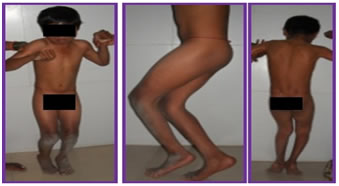
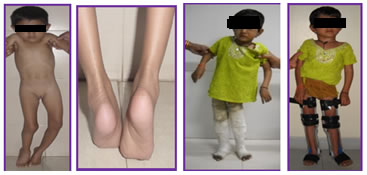
According to Modified Functional walking scale, pre-operatively 9 children (26.47%) were in non-walking grade (Level 1 and 2), 22(64.70%) in therapeutic walkers (level 3 and 4), 3(8.82%) in household walking grade (level 5) and 0 in community walking grade (level 6 to 10). Post-operatively all children improved and moved out of non-walking grade. 19 patients (55.87%) progressed to be therapeutic walkers (level 3 and 4), 11 patients (32.35%) to household walking grade (level 5) and 4 patients (11.76%) to community walkers (level 6 to 10). None improved beyond level 6. Thus maximum children (33 out of 34) were found to have improvement, which was significant, in their Modified Functional walking scale after Vulpius recession surgery. Out of 34 patients, 26 patients (76.47%) required some type of support for mobilization pre-operatively while only 8 patients (23.52%) were able to ambulate without any external support, whereas post-operatively only 11 patients(32.35%) required support for mobilisation while 23 patients (67.65%) could mobilize without external support. Thus, dependency on external support for mobilisation was reduced in patients who underwent Vulpius recession surgery. Figure 1: Figure 2: DISCUSSION Dynamic derangement in the gait of children with diplegia is caused by spasm and imbalance in the muscles of both the leg. Although the hip and knee and their related muscles are also affected, the deformity which has the greatest effect on gait and mobility is an equinus deformity of the ankle. However, equinus deformity is often more ‘apparent’ than ‘real’. Spasticity and flexion contractures at the hip and knee may dictate an equinus posture at the ankle when there is no calf contracture because of the need to achieve sagittal plane balance. This is a biomechanical response to control the direction of the ground reaction vector in relation to the centres of joint rotation. Hence, we first distinguished between true equinus and apparent equinus. We have used observational gait analysis to study the effect of gastrosoleus recession using Vulpius technique as it can be easily performed in clinical setting and doesn’t require expensive set up necessary for instrumented gait analysis. In a previous study, they have found observational gait analysis to have good inter- and intra-observer repeatability.6 Vulpius technique is the oldest technique of gastrosoleus recession to correct equinus deformity in cerebral palsy. It was first used in 1913 and since then it has proved to be a simple and safe procedure requiring a small incision, no need of tendon suturing, no wound healing problems, delayed or non-union of tendon and when done appropriately over-lengthening (resulting in calcaneus deformity) can be avoided. In spastic type of cerebral palsy, spasticity interferes with normal movements. Recession at the musculotendinous junction decreases spasticity. Hence, we have used the Vulpius procedure to correct equinus deformities and study clinically its effect on gait. Aponeurotic lengthening are inherently more stable than tendon lengthening’s. This is probably the reason why in our study the Vulpius operation was associated with no case of overlengthening, despite the fact that it is not a selective gastrocnemius procedure. We performed the Silverskiold test7 on all patients but irrespective of its result we performed Vulpius procedure on all patients as it is equally applicable whether the contracture is in gastrocnemius muscle, the soleus muscle, or both.8 After evaluating every patient pre-operatively, we performed single event surgery at single or multiple levels (hip/knee/ankle) as per the requirement of the patient. Partial neurectomy of the gastrosoleus was not done at the time of gastrosoleus recession because of its liability to produce calcaneus.9 We agree with Gaines and Ford that over-correction with subsequent calcaneus gait is a disastrous complication.10 It is more satisfactory to do a further operation to correct recurrent equinus than to risk a calcaneus gait or even calcaneus deformity. There is good experimental evidence to suggest that the calf muscle responds by adding sarcomeres and gradually becomes anatomically too long and biomechanically incompetent.11,13 In time, a calcaneus gait may progress to a calcaneus deformity.14 Management of calcaneus deformity is much more difficult and there is a greater need to avoid it.14,15 Assessment of outcome after surgery for calf lengthening is difficult. Sagittal plane kinematics and kinetics of the ankle are the most sensitive outcome measures, as both under- and overcorrection can be readily identified. Sharrard and Bernsteinshowed a 23% recurrence rate after tendocalcaneus elongation and 15% after gastrocnemius recession.9 In our study we had a recurrence rate of 2.95%. We used ‘Foot part’ of Physician Rating scale to assess change in equinus post-operatively. 33 patients had an improved (heel-strike or flat foot) gait at one-year follow-up. We measured maximum passive dorsiflexion of the foot in all children before and after surgery. All children except one had maximum passive dorsiflexion between 00-200 indicating improvement in ankle range of motion and its function during gait. Khan MA used the functional walking scale. In his study all the children improved and moved out of the non-walking grade; 18 (21.2%) progressed to be therapeutic walkers (level 3 and 4), 39 (45.9%) to household walkers (level 5) and 28 to community walkers (level 6 to 10). None improved beyond level 8 and only six (7%) reached this level.16 In our study too, post-operatively all children improved and moved out of non-walking grade. 19 patients (55.87%) progressed to be therapeutic walkers (level 3 and 4), 11 patients (32.35%) to household walking grade (level 5) and 4 patients (11.76%) to community walkers (level 6 to 10). None improved beyond level 6. Khot A et al studied 16 cerebral palsy children. At the time of entry to the study, 5 children were GMFCS level III and 11 were level IV. A non-significant improvement in GMFCS levels was found after intervention.17 Ma et al noticed that two patients out of 19 were improved by one GMFCS level. The GMFCS is believed to be stable over time. In our study, no patient had change in the GMFCS grade.18Saraph V et al used Modified Ashworth Score for grading spasticity and found decrease in spasticity following surgery.19In our study too, spasticity was decreased when evaluated by Modified Ashworth score. Knee-ankle-foot orthotic braces or ankle-foot orthotic braces were prescribed based on post operative assessment of gait in each child. Patients with cerebral palsy require prolonged rehabilitation and follow-up after surgery. Stretching excercises have not shown evidence of reducing the recurrence rate.It is rare that a child or adult sleeps with his/her feet in the neutral position or in dorsiflexion. Most commonly, regardless of the position of the child, the ankles tend to be in some plantar flexion. The braces are intended to avoid maintaining this position for the night time, approximately 1/3rd of the child’s day. We gave night splinting after surgery for correct of equinus contracture from 6 months until completion of growth. In our series, 33 out of 34 (97.05%) patients were satisfied with the results.
CONCLUSION Gastrosoleus recession by Vulpius method is an effective procedure in treatment of spastic diplegia type cerebral palsy patients with equinus. Patient had good improvement indicated by need for brace, support for mobilization, maximum passive dorsiflexion of ankle, Physician rating scale, Modified functional walking scale, Modified Ashworth scale with minimal complications.
Limitations of the study There are, however, a number of limitations to this study. It describes a very specific group of children with bilateral spastic cerebral palsy treated with relatively limited surgery in a specialist centre and the conclusions may not be generalized to other age groups or other surgical approaches. The group size is small, and the length of follow-up of the patients in the operative group was limited i.e one year. We assessed the effect of surgery on gait and functional abilities but not on health-related quality of life for each child, and future prospective studies will be needed for this. The goals and outcome of surgery were limited but are still valid.
REFERENCES
|
|
 This work is licensed under a Creative Commons Attribution-NonCommercial 4.0 International License.