 Home
HomeOfficial Journals By StatPerson Publication
|
Table of Content - Volume 9 Issue 2 - February 2019
Complications of distal humerus fractures treated with open reduction and internal fixation with bicolumnar plating by extensor mechanism sparing paratricipital approach
Rajesh K Ambulgekar1, Kapil R Saoji2*
1Professor and Head, 23rd year Resident, Department of Orthopaedics, Dr Shankarrao Chavan Government College and Hospital Nanded, Maharashtra, INDIA. Email: drkapil.saoji@gmail.com
Abstract Background: Distal articular humerus fractures when managed conservatively has high chances of incongruous joint, non-union, malunion, and stiff elbow. The paratricipital approach have several advantages but it has disadvantage also. Aim: To evaluate the complications of fracture of distal humerus in adults treated with open reduction and internal fixation with bicolumnar plating by extensor mechanism sparing paratricipital approach. Material and Methods: A total of 30 patients with distal end humerus fracture were treated with open reduction and internal fixation with bicolumnar plating by extensor mechanism sparing paratricipital approach and followed up for complications. Results: Complications occurred in 6 patients (20%). Three patients had stiffness (elbow ROM <1000) but all had functional range of motion. Two patients had superficial infection which was subsided with oral antibiotics. One patient had screw loosening and back out. Conclusion: Open reduction and internal fixation with bicolumnar plating by extensor mechanism sparing paratricipital approachavoids complications associated witholecranon osteotomy. It gives in excellent healing and better outcome. Key Word: distal humerus fracture, Para-tricepital approach, complications
INTRODUCTION The management of distal humeral fractures has evolved over the years from non-operative treatments, such as the so called bag- of -bones technique to operative treatments.1 Following the conservative approach chances of incongruous joint, non-union, malunion, and stiff elbow are very high. Therefore, most condemned conservative management in all type of fractures, and advised surgical management. Distal articular humerus fractures are preferably treated by open reduction and internal fixation.2 Multiple exposures such as olecranon osteotomy approach and dissociation of the triceps from the olecranon have been described. But these are not free of complications and that complications necessitate more surgery and predispose to infections.3To avoid these complications an extensor mechanism sparing paratricipital posterior approach to Distal Humerus through midline posterior incision was suggested by Schildhauer et al.4 The paratricipital approach have several advantages, complications of olecranon osteotomy can be avoided, triceps tendon insertion not disrupted, allows early range of motion. This approach also preserves innervations and blood supply of anconeus muscle5,6 which provides dynamic postero-lateral stability of elbow. The disadvantage of paratricepital approach is the limited visualisation of articular surface of distal humerus, therefore this approach is usually inadequate for fixation of type C3 fractures.7,8 The purpose of this study is to evaluate the complications of fracture of distal humerus in adults treated with open reduction and internal fixation with bicolumnar plating by extensor mechanism sparing paratricipital approach.
MATERIAL AND METHODS In this prospective study, patients with distal end humerus fracture were treated with open reduction and internal fixation with bicolumnar plating by extensor mechanism sparing paratricipital approach. The study was conducted in Department of Orthopedics of a Tertiary care hospital over a period of two years. Informed written consent was taken for all patients and approval from Institutional Ethical Committee was obtained prior to the commencement of the study. Inclusion Criteria
Exclusion Criteria
METHODOLOGY Detailed history was taken from the patient regarding the etiology of the fracture, associated injuries, general examination, physical examination of the corresponding shoulder, elbow and wrist joints was carried out. Investigations were done in the form of elbow X-rays (AP and lateral views; both oblique views if required) and were evaluated. Fractures were classified based on the AO classification. Primary management was done and fracture immobilization in above elbow slab upto mid-arm level and the patient was shifted to ward with elevation of the affected upper limb. Patients included in the study were treated with open reduction and internal fixation with bi-columnar plating by Extensor Mechanism sparing paratricipital approach, and were assessed intra-operatively for blood loss, fracture reduction and articular continuity under the image intensifier (C-arm). Post-operatively, patients were followed up post-operatively at 1 month, 2 months, 6 months after treatment, and were evaluated clinically by Mayo Elbow Performance Score at each follow-up visit, along with X-rays (AP and lateral views). Secondary outcome measures used consisted of Elbowpain, Active and passive ROM (flexion, extension) of both elbow joints using a universal goniometer, Disability inperforming daily activity. Complications such as infection, neurovascular compromise, stiffness, subsequent or secondary intervention, arthritis were looked. X-rays (antero-posterior and lateral views) were evaluated on every follow up for the signs of fracture healing and hardware failure or any other complications. Union was defined as the presence of bridging callus or the disappearance of the fracture line on three of four cortices seen on the anteroposterior and lateral radiographs.
Figure 1 Figure 1: Temporary stabilization using K-wires Figure 2: The lateral and medial column plates Figure 3: Drain kept in situ and closure is done in layers
RESULTS Out of the 30 cases, majority of case i.e. 17 (56.67%) were in the age group of >30 years. The minimum age of 13 years and maximum of 72 years with mean age of 38.77 years. Out of 30 patients 17 patients (56.67 %) were males and 13 patients (43.33 %) were females in our study. Road traffic accidents account for 18 (60%) of the cases as a cause and remaining 12 (40%) cases had history of fall or slip. Right side has a marginal high predominance 20 (66.67%) than left side 10 (33.33%). The common fracture type (AO classification) we accounted in our study were Type A2 which was in 9 patients (30 %) and Type A3 which was also in 6 patients (20 %), Type B1 in 3 patients(10%), Type B2 in 3 patients(10%), Type B3 in 4 patients(13.33%), Type C1 in 2 patients(6.67%) and Type C2 in 3 patents(10%).By using ANOVA test we found no significant difference between mean ROM with respect to AO fracture type.
Table 1: Correlation between AO type and ROM (6 Month)
In our study complications occurred in 6 patients (20%). 3 patients had stiffness (elbow ROM <1000) but all had functional range of motion, so no intervention was done. 2 patients had superficial infection which was subsided with oral antibiotics within 14 days. 1 patient had screw loosening and back out which became palpable under the skin, the screw was removed under short General anesthesia and patient became pain free again. Apart from these no other complications were observed in our study. Table 2: Complications
DISCUSSION The common fracture type (AO classification) we accounted in our study were Type A2 which was in 9 patients (30%) followed by type A3 which was also in 6 patients (20%). This distribution was comparable to the study by Mondal et al where 50% fractures were Type A, 33.33% fractures were Type B, 10% of fractures were Type C1, 6.66% of fractures were Type C2.9Patel et al accounted for 9 cases (22.5%) of type A, 5 cases (12.5%) of type B, 26 cases (65%) of fractures of type C.10 Previous investigators of triceps-splitting or peeling approaches had postulated a negative effect on muscle strength on the basis of the potential for weakened reattachment, direct muscle injury with resultant fibrosis, and injury to intramuscular nerve branches. Our results compared favorably with other studies utilizing different approaches, as this approach maintained the triceps attachment to the olecranon, eliminated the need for triceps repair and protection postoperatively, allowed active range of motion in the injured elbow. Restoration of the articular surface was the most important step followed by stabilization of the largest columnar fragments. Several options were available for fixation, these include the use of Y-shaped plates, reconstruction plates, LC-DCP, single plates, pre-contoured locking distal humerus plates. The aim was to achieve the stable reconstruct. There was still debate going on about, which plate positions provide optimal stability for distal humerus fractures. Jacobson SR et al. tested five different distal humerus plating constructs in cadaveric specimens. They concluded that a medially applied 3.5 mm reconstruction plate combined with a posterolateral (orthogonal) 3.5-mm dynamic compression plate provided the greatest sagittal plane stability, and equivalent frontal plane and torsion stability, when compared with other constructs, which included parallel and triple plating.11Mardanpour K et al also used bilateral plates, which were pre bended according to morphology, applied on medial and posterolateral sides of humerus at 45 to 900.12 In our study we placed the lateral column plate dorsally curving around the capitulum and lateral condyle and the medial column plate on the medial surface of the medial supracondylar pillar. In this position the planes of 2 plates formed an angle of approximately 90 degrees to each other. This provided both antero-posterior and medio-lateral stability. We found this orthogonal plate construct provided good stability, which was checked intra operatively, by allowing elbow through full range of motion, and on regular follow ups. In our study complications occurred in 6 patients (20%). Three patients had stiffness (elbow ROM <1000) but all had functional range of motion, so no intervention was done. Two patients had superficial infection which was subsided with oral antibiotics within 14 days. One patient had screw loosening and back out which became palpable under the skin, the screw was removed under short General anesthesia and patient became pain free again. Apart from these no other complications were observed in our study. Reported complication from other studies includes ulnar neuropathy, malunion/ nonunion/ delayed union, implant failure, contracture. In the study of Mondal et al, symptomatic hardware was seen in 3 patients, superficial skin infection was seen in 13.33%, tourniquet palsy was seen in 1 case (3.33%), ulnar nerve neuropraxia was seen in 2 cases.9 In the study of Ali et al, there was one deep infection, no implant failure, neurovascular deficit or nonunion.13 In the study of Patel et al, complications like superficial infection in 6 (15%) patients, non-union in 3 (7.5%) patient, palpable implants in 4 (10%) patients were noted.10 In the study of Yadav et al two patients had experienced ulnar neuropathy, superficial infection was detected in two patients, no evidence of heterotopic ossification.14 Stiffness was a common complication of fractures of the distal humerus and was most often caused by inadequate post-operative rehabilitation. Early active motion permitted by this approach, as continuity of the triceps was maintained, could minimized formation of intraarticular adhesions and periarticular fibrosis that may negatively affect the range of elbow motion. There were certain limitations of this study as well. A larger sample population needed to be studied to reduce the "type II" or beta error of the study. A better method of randomization should have been adopted. The patients could not be followed up for a longer period.
REFERENCES
|
|
|||||||||||||||||||||||||||||||||||||||||||||||||||||||||||||||||||||||||
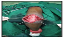
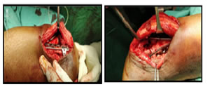 Figure 2 Figure 3
 This work is licensed under a Creative Commons Attribution-NonCommercial 4.0 International License.