 Home
Home
Official Journals By StatPerson Publication
|
Table of Content - Volume 11 Issue 2 - August 2019
A comparative study of clinical presentation of pancytopenia and bicytopenia in hospitalized children
Chaure Varsha P1, Khaire P B2*, Mundada Smita3
1JR III, 2,3Associate Professor, Department of Pediatrics, Government Medical College, Aurangabad, Maharashtra, INDIA. Email: pmorekhaire@rediffmail.com
Abstract Background: Pancytopenia and Bicytopenia are results of failure of production of hematopoietic progenitors in bone marrow, malignant cell infiltration, antibody mediated bone marrow suppression, and ineffective hematopoiesis and peripheral sequestration of blood cell in overactive reticuloendothelial tissue. Aims and objective: To compare Clinical Presentation of Pancytopenia and Bicytopenia in Hospitalized Children. Materials and Methods: In the present study all hospitalized children with pancytopenia / bicytopenia between 1months -12 yrs of age observed on Complete blood count and peripheral smear during the study period were included in the study. Thus total 208 cases were enrolled in the study out of them 128 patients were of bicytopenia while 80 patients were having pancytopenia. Selected children were assessed after written informed consent from parents and enrolled in study. Complete demographic data which includes bio-data of patient, complete history, clinical examination and investigations as per proforma was recorded and maintained. 2 ml of blood was collected by venipuncture under aseptic precaution in a dry bulb containing ethylene diamine tetra acetic acid (EDTA) anticoagulant. Then sample was put in the automated haemotology analyzer (medonic) and hemoglobin, total leucocyte count, platelate counts were noted. Peripheral smear prepared and stained by fields A and fields B (romanowsky). Microscopic examination was done systematically under low, high and oil immersion for RBC morphology, Total leucocyte count and platelate count and this data was noted. Results: Amongst 208 cases, Bicytopenia (128) was more common than Pancytopenia (80). The most common age group affected in bicytopenia was 1 month –12 months (35.1%) and in pancytopenia, it was 61months-144 months (36.3%).In sex distribution, bicytopenia had M:F ratio of 0.8:1 and Pancytopenia had 1.05:1 Fever was the most frequent symptom found in both bicytopenia (97%) as well as pancytopenia (87.5%). In general examination, Pallor was the most common clinical sign found in bicytopenia (77.34%) and pancytopenia (83.7%). Etiologically, infectious causes were more common than non infectious in both bicytopenia as well as pancytopenia. Conclusion: Thus we conclude that the most common age group affected in bicytopenia was 1 month – 12 months and in pancytopenia, it was 61months–144 months. Fever, cough and lethargy were the most frequent symptom found in both bicytopenia and Pancytopenia patients. Pallor, hematomegaly and rash were the most common clinical sign observed in bicytopenia and Pancytopenia as well. This study showed that commonest etiology of both bicytopenia and Pancytopenia was infections. Key Words: Pancytopenia, bicytopenia, Clinical Presentation
INTRODUCTION Pancytopenia is an important clinico-hematological entity encounter in our day to day clinical practice. There are varying trends in its clinical pattern, treatment modalities and outcome.1 Pancytopenia refers to a reduction below normal value of all three peripheral blood lines, leucocytes, Platelates, and erythrocytes. There are three general categories as per bone marrow findings which are hypocellular, hypercellular and bone marrow infiltration.2,3 While Bicytopenia is defined as in reduction of any two parameters from the complete blood count.3,4,5 Pancytopenia and Bicytopenia are results of failure of production of hematopoietic progenitors in bone marrow, malignant cell infiltration, antibody mediated bone marrow suppression, and ineffective hematopoiesis and peripheral sequestration of blood cell in overactive reticuloendothelial tissue. Common conditions associated with pancytopenia and bicytopenia are aplastic anemia, megaloblastic anemia, and acute leukemia. Common clinical feature of pancytopenia and bicytopenia are fever, petechial rash, bleeding manifestation and hepatosplenomegaly.6 This study is carried out to identify the causes of pancytopenia and bicytopenia, to find out frequency of different causes and to determine the incidence of pancytopenia and bicytopenia in relation to sex, age as well as to compare our findings with those of other similar studies from other part of the world. Pancytopenia is common hematological problem with an extensive differential diagnosis and still the diagnostic approach to pancytopenia remains undefined in our country. The cause of pancytopenia and bicytopenia is not well defined. So this study will evaluate the various causes and the data will help in planning the diagnostic and therapeutic approach in patient with pancytopenia and bicytopenia.1
MATERIALS AND METHODS The present cross sectional study was conducted in the Department of Paediatrics, of Govt. Medical Collage and Hospital during November 2016 to November 2018. All hospitalized children with pancytopenia / bicytopenia between 1months - 12 years of age observed on Complete blood count and peripheral smear during the study period were included in the study. Thus total 208 cases were enrolled in the study out of them 128 patients were of bicytopenia while 80 patients were having pancytopenia.Pancytopenia refers to a reduction below normal value of all three peripheral blood lines, leucocytes < 4000/cmm, platelets < 1lac/cmm and hemoglobin <10 gm/dl.2,4,5 Bicytopenia refers to reduction of any two parameters from the peripheral bloodlines, Hemoglobin <10 gm/dl, leucocytes <4000/cmm, platelets <1lac/cmm4,5.Children with lukaemia who are already diagnosed and on chemotherapy or already diagnosed children with aplastic /hemolytic anemia with recent blood transfusion or children on immunosuppressant therapy were excluded from the study. Children who fulfilled selection criteria were assessed after written informed consent from parents and enrolled in study. Complete demographic data which includes bio-data of patient, complete history, clinical examination and investigations as per proforma was recorded and maintained 2 ml of blood was collected by venipuncture under aseptic precaution in a dry bulb containing ethylene diamine tetra acetic acid (EDTA) anticoagulant. Then sample was put in the automated haemotology analyzer (medonic) and hemoglobin, total leucocyte count, platelate counts were noted. Peripheral smear prepared and stained by fields A and fields B (romanowsky). Microscopic examination was done systematically under low, high and oil immersion for RBC morphology, Total leucocyte count and platelate count and this data was noted. The collected data was processed and analysed by Microsoft excel software and qualitative (chi square test) statistical tests were applied.
RESULTS Table 1: Distributions according to age and sex in Bicytopenia and Pancytopenia
Total number of enrolled patients as per the inclusion and exclusion criteria were 208, among that 128 (61.53%) patients were bicytopenia while 80 (38%) patients were having pancytopenia. Among the 128 children with Bicytopenia, maximum number of patients (35.15%) were in the age group between 1m to 12 months, followed by 61 months to 144 months (32%) and patients observed in the age group between 13 months to 60 months (31.5%). Among the 80 children with Pancytopenia, maximum number of patients (36.2%) were in the age group between 61 months to 144 months, followed by 13 months to 60 months age group (33.7%) and between 1 month to 12months age group (30%). The difference observed in the distribution of age in bicytopenia and pancytopenia was statistically insignificant. Out of the 128 children with Bicytopenia, 45% were male and 64% were female with male to female ratio of 0.8:1. In pancytopenia among 80 patients, 51% were male and 35% were female, with male to female ratio of 1.05: 1. The distribution of male and female in bicytopenia and pancytopenia group was statistically insignificant (p value >0.05)
Table 2: Distributions according to symptoms in Bicytopenia and Pancytopenia
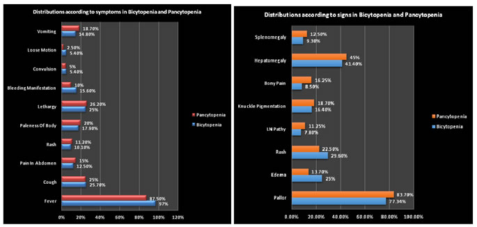
In Bicytopenia, fever (97%) was the most common symptom followed by cough (25.7%), lethargy (25), abdominal pain (12.5%), petechial rashes (10.1%), paleness of body (17.9%) and bleeding manifestation (15.6%). In Pancytopenia, Fever (87.5%) was the most common symptoms followed by lethargy in 26.2%, cough in 25%. vomiting in 18.7%, paleness of body in 20%, bleeding manifestation in 10%, rash in 11.2%, and vomiting in 18.7%. The difference observed in the occurrence of symptoms in Bicytopenia and Pancytopenia patients was statistically non significant.
Figure 1: Distributions according to symptoms in Bicytopenia and Figure 2: Distributions according tosigns in Bicytopenia and Pancytopania Pancytopania Table 3: Distributions according to signs in Bicytopenia and Pancytopenia
In Bicytopenia, pallor (77.34%) was the most common sign, followed by hepatomegaly (41.4%), rash (29.6%), edema (25%), knuckle pigmentation (16.4%), bony pain (8.5%) and splenomegaly (9.3%). In Pancytopenia, pallor (83.7%) was the most common clinical sign, followed by hepatomegaly (45%), rash (22.5%). Bony pain was seen in 16.25%, edema in 13.7%, splenomegaly in 12.5% patients. It was seen that the difference observed in the presence of sign in Bicytopenia and Pancytopenia patients was statistically non significant. Table 4: Distributions according to etiology of Bicytopenia and Pancytopenia
In Bicytopenia, more number of cases were of infectious etiology (61%) than non infectious (38.2%). Also in Pancytopenia, infectious cases (57.5%) were more commonly observed than non infectious (42.5%). Over all infectious cases (60%) were more commonly observed than non infectious (39.9%).
DISCUSSION Pancytopenia is defined as presence of anaemia, thrombocytopenia and leucopenia. Pancytopenia is a common occurrence in pediatric patients. Patients usually present with symptoms due to anaemia, leucopenia or thrombocytopenia. Identification of etiology of pancytopenia is crucial as the underlying etio-pathology determines the management and prognosis of the patients. There is only very few data reported on clinical and etiological spectrum in children with Bicytopenia. Bicytopenia can be due to transient viral illness or life threatening malignancies. Different population groups show difference in disease presentations based on their genetic pattern, prevalent infections in that location, and also their nutritional status. Etiological profile also shows difference with different researches. In present study most common affected age group among bicytopenia patients was between 1month to 12 months (35.1%) followed by age group between 61 months to 144 months (32%). Similar age distribution in bicytopenia, was noted in study done by Rehman et al (2017)3. The age group criteria was different in, Saadia et al(2015)6 it was 10yr- 30 yr (36%) while in Muddasir et al(2014)4, it was 61months -144 months (39%) which was the second most common age group in present study. In present study, in pancytopenia it was observed that age group between 61 months to 144 months was the most commonly affected 29(36.3%) followed by between13 months to 60 months (33.7%). Similar age distribution of pancytopenia was noted in Tufail et al (2017)8. The age group criteria was different in Anwar et al(2013)6, It was 6months – 5 yrs and Ghanshyam et al(2016)9, it was between 12 months - 60months.Out of the 128 children with Bicytopenia, 45% were male and 64% were female with male to female ratio of 0.8:1. While Naseem et al(2011)5 found, 64% male and 35% were females and male to female ratio was 1.8:1. Muddasir et al(2014)4 found, 53% male and 46% females and male to female ratio was 1.2:1. Zahide et al(2014)13 found 57% male and 42.9% female and male to female ratio was 1.3:1. Siraj et al(2017)10 found 59.1% female and 40.99%. In pancytopenia among 80 patients, 51% were male and 35% were female, with male to female ratio of 1.05:1. Chate et al(2012)11 found that male (56%) were more than female (43.3%) with male to female ratio 1.3:1. Anwar et al(2013)6 also found that male (64.8%) more than female with male to female ratio were 1.8:1. Naseem et al(2011)5 found that 61.1% were male and 38.1% female. In contrary Dubey et al (2016)12 found that females (53%) were more than male (47%) with male to female ratio 0.88:1. The distribution of male and female in bicytopenia and pancytopenia group was statistically insignificant (p value >0.05). In present study, in bicytopenia patients fever (97%) was the most common symptom followed by cough (25.7%), lethargy (25%), paleness of body (17%), bleeding (15.6%) and vomiting (14.8%). Naseem et al(2011)5 in their study found that fever (69.25%) was the most common symptom followed by petechial rash (17.9%), bleeding (12.3%) and bony pain (9.5%). Muddasir et al(2014)4 also found that fever (82.9%) was the most common symptom followed by anorexia (21%) patients, bleeding (32%), lethargy (16.2%), irritability (4.8%) and Pica (1.9%). Zahide et al(2014)13 in their study observed that fever (67.8%) was the most common symptom followed by exhaustion (14.2%) and petechial rash (14.2%). Shilpi et al(2018)14 also found that fever (41.7%) was the most common symptoms followed by generalized weakness (27%), bleeding (12%) and petechial rash (6.2%). Thus in bicytopenia, like in present study similar clinical symptomatological profile was noted in studies done by Naseem et al(2011)5, Zahide et al(2014)13, Shilpi et al(2018)14 as well as Muddasir et al(2014)4. Among the pancytopenia patients fever (87.5%) was the most common presentation followed by lethargy (26.2%), cough (25%), paleness of the body (20%), and vomiting (18.7%). Anwar et al(2013)6 also found fever (62.8%) as the was the most common symptom followed by bruises (62.4%), petechial rash (19.5%), hematuria (12.1%) and bleeding gum (24.3%). Chate el al (2015)17 in their study found that fever (67.3%) was the most common symptom followed by bleeding (54.3%), weight loss (23.9%), and bone pain (4.3%). Tufail et al(2017)15, reported that fever (92%) was the most common symptom followed by joint pain (29.6%) followed by bleeding (20%).In present study, among the bicytopenia patients it was found that pallor (77.34%) was the most common sign, followed by hepatomegaly (41.4%), rash (29.6%), edema (25%), knuckle pigmentation (16.4%), bony pain (8.5%) and splenomegaly (9.3%). Naseem et al(2011)5 found that hepatomegaly (69.2%) was the most frequent clinical sign followed by splenomegaly (60.9%), lymphade- nopathy (41.9%), pallor (40%) and bony pain (9.5%). Muddasir et al(2014)4 found that hepatomegaly (27.6%) was most frequent sign followed by splenomegaly (4.8%) and lymphadenopathy (5.7%) Zahide et al(2014)13 found that hepatomegaly (10.7%) was the most frequent clinical sign followed by splenomegaly (3.5%) and short stature (3.5%). Shilpi et al(2018)14 found that splenomegaly (41.7%) was the most frequent sign observed followed by hepatomegaly (27%) and lymphadenopathy (25%). In present study, among pancytopenia patients pallor (83.7%) was the most common clinical sign followed by hepatomegaly (45%) and splenomegaly (12.5%). Other clinical signs were rash (22%), knuckle hyper pigmentation 15(18.7%), and bony pain 13(16.3%). Similarly Anwar et al(2013)6 found that pallor (82.9%) as the most common clinical sign. Chhabra et al(2012)16 found pallor (64.8%), hepatomegaly (59.3%) and splenomegaly (57.1%) as the common clinical signs. Chate et al(2015)17 found that pallor (63%) and hepatomegaly (56.5%) as the most common clinical signs followed by splenomegaly (41.3%) and other clinical signs were lymphadenopathy (15.2%). Tufail et al(2017)15 also found that pallor (83.2%) and hepatosplenomegaly (64.8%) as the most common signs followed by petechiae (42.4%).In Bicytopenia, more number of cases was infectious (61%) than non infectious (38.2%). Similar findings were also reported by Saadiya et al (2015)18 Zahide et al (2014)13 and Rehman et al (2015)19. Also in Pancytopenia, infectious cases (57.5%) were more commonly observed than non infectious (42.5%). Over all infectious cases (60%) were more commonly observed than non infectious (39.9%). Similar observation were also reported by, Anwar et al (2013)6, Tufail et al (2017)15, Chabra et al (2012)16, Chate et al (2015)17 and Bhatnagar et al (2005)20.
CONCLUSION Thus we conclude that the most common age group affected in bicytopenia was 1 month – 12 months and in pancytopenia, it was 61months–144 months. Fever, cough and lethargy were the most frequent symptom found in both bicytopenia and Pancytopenia patients. Pallor, hematomegaly and rash were the most common clinical sign observed in bicytopenia and Pancytopenia as well. This study showed that commonest etiology of both bicytopenia and Pancytopenia was infections.
REFERENCES
|
|
||||||||||||||||||||||||||||||||||||||||||||||||||||||||||||||||||||||||||||||||||||||||||||||||||||||||||||||||||||||||||||||||||||||||||||||
 This work is licensed under a Creative Commons Attribution-NonCommercial 4.0 International License.