 Home
Home
Official Journals By StatPerson Publication
|
Table of Content - Volume 12 Issue 2 - November 2019
Neonatal morbidity and mortality in post-term pregnancy in civil Hospital affiliated to B J Medical College, Ahmedabad, Gujrat
Manisha Ghoniya1*, Anuya Chauhan2
1Resident, 2Assiatant Professor, Department of Paediatrics, BJ Medical College, Ahmedabad, Gujrat, INDIA. Email: manishaj.khunt@gmail.com
Abstract Background: Post-term pregnancy is defined as gestation that extends beyond 42weeks.The relative perinatal mortality is higher in post-term delivery compared with delivery at term and has been associated with an increased frequency of neonatal morbidity (meconium aspiration, fetal distress, asphyxia in the neonatal period, pneumonia, malformations, macrosomia and fetal birth injury) and maternal complications (cesarean section, postpartum hemorrhage, labor dysfunction and obstetric trauma). Although it is known that these risks are increased in post-term pregnancies, what has received less attention is whether and to what extent these risks increase before 42 weeks of gestation. Objective: To study Neonatal outcome in terms of morbidity and mortality in cases of postdate pregnancy (> 40 weeks) and its comparison with that of term pregnancies (37-40 weeks). Study Design: This was a Prospective Observational Case control study, conducted at Pediatric department, Civil Hospital affiliated to B.J. Medical College, Ahmedabad from September 2011 to August 2013. Result: A total of 200 babies were analyzed; of these, 81% case 40 to 41weaks of gestational age,16 %cases were 41 to 42weaks gestational age, and 3% cases were of more than 42 weeks gestational age.27 % cases have history of oligohyramniosis,41% cases had LSCS deliveries,3% cases had APGAR Score less than 4, meconium stained liquor seen in 25% cases of 41 to 42 weeks gestational group babies. Neonatal morbidity in form of IUGR in 2%, MAS in 7%, TTN in 8%, Birth asphyxia in 3%, PMS in 3%, of cases observed. Neonatal mortality was 7% in post date babies. Maternal complication in form of Post partumhemorrage in 5%, 3 -4 degree perineal tear in 2%, and cervical tear in 3% cases were observed. Conclusion: One should consider postdate pregnancy as a high-risk condition. Even uncomplicated post-date pregnancy is associated with increased rates of obstetric and neonatal interventions in terms of caesarean section and NICU admissions. Management of women with postdate pregnancy should be individualized taking into consideration the amount of liquor and the findings of sonography and NST there by demanding early detection and proper planning. Considering the above-mentioned adverse perinatal outcome. Key Word: neonatal outcome, post term pregnancy, postmaturity syndrome
INTRODUCTION There is definite clinical entity, “POST DATISM”, which is associated with high foetomaternal risk. As the gestational age increases after 37 completed weeks, the incidence of perinatal morbidity, mortality and maternal peripartum complications also increases with each passing week of gestation.1 Postdate pregnancy is also associated with increased costs related to antenatal fetal monitoring and induction of labour and can be a source of significant anxiety for the pregnant woman. The emerging evidence demonstrates that the incidence of complications associated with postdate pregnancy also increase from 40 to 42 weeks, including rate of induction, length of labour, prolonged second stage of labour, instrumental delivery, caesarean delivery, postpartum haemorrhage, cephalopelvic disproportion, cervical rupture, severe perineal injuries, dystocia and puerperal sepsis.2,3,4 Foetal complications like risks of meconium aspiration, asphyxia before, during and after delivery, macrosomia, foetal birth injury, foetal septicaemia, rate of non-reassuring foetal heart rate or foetal death during delivery, also significantly increases.2,3 In 1954 Clifford described a syndrome found in infant born after the expected date of delivery which in many aspects resembled intrauterine growth retardation. There was often thick meconium staining of the amniotic fluid and signs of fetal distress in these postmature infants.5Despite intensive research, management of postdate pregnancy is still controversial and differs, not only among different countries and hospital, but also among different clinicians in the same hospital. This subject was chosen because while it is well established that these risks increases in post term pregnancy, what is less proven is, if these risks increase prior to 42 weeks gestation. In present day where small family norm is opted, child's survival is very crucial. This is a critical issue in our developing country where extensive family planning programs are going on. Postdate pregnancy is one that exceeds 40 weeks (280 days) or more from the first day of onset of last menstrual period. Post term pregnancy is one that exceeds 42 weeks (294 days) or more from the first day of the last menstrual period.
AIMS AND OBJECTIVES To study foetal outcome in terms of morbidity and mortality in cases of postdate pregnancy (> 40 weeks) and its comparison with that of term pregnancies (37-40 weeks).
METHODS This was a Prospective Observational Case control study, was conducted at Pediatric department, Civil Hospital affiliated to B.J. Medical College, Ahmedabad from September 2011 to August 2013. A total 200 newborns who were delivered at Civil Hospital affiliated to B.J. Medical College, Ahmedabad were studied. Multiple births, babies with congenital malformations or inaccurate gestational age were excluded. For each delivery, a trained field worker completed a questionnaire, which included pertinent, social, gynecological, obstetric, perinatal and baseline maternal medical data. All babies were followed up until discharge. Study subjects were further divided in two groups according to gestational age. Cases: It included total hundred Newborn with gestational age more than 40 weeks. Controls: It included total hundred Newborns with gestational age between 37 weeks and 40 weeks. Gestational age was calculated by dates (when known) oral tentatively by Ballard Score, or prenatal ultrasound. We examined the following outcomes: maternal vaginal bleeding, chorioamnionitis, emergency cesarean delivery, elective cesarean delivery, 5 min Apgar score less than 4, acute fetal distress, macrosomia, meconium aspiration syndrome, admission to the neonatal intensive care or step-down unit, stillbirth and neonatal death. Maternal Outcome in terms of Mode of delivery — Vaginal or LSCS or instrumental delivery, cervical or vaginal tear/lacerations, PPH were noted. The neonatal outcome was correlated with degree of postdatism and it was compared with controls. Perinatal mortality, perinatal morbidity, Apgar score, incidence of respiratory distress, meconium aspiration syndrome, birth asphyxia, septicaemia, admission in NICU were noted. Statistical Analysis: Qualitative data was expressed as number (frequency) and Chi-square test was used as the test of significance. Quantitative data was expressed as mean and standard deviation, and independent t-test was applied for normally distributed data and Mann-Whitney test for non-normally distributed data. All analysis was carried out using Statistical Package for Social Sciences (SPSS) version 22.0 and p values <0.05 were marked as the level of Significance.
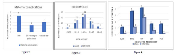
RESULTS On basis of pre-decided inclusion and exclusion criteria, a total of 200 newborns who were delivered in labour room /ward were enrolled for the study. Table 1 shows distribution of maternal age, out of 100 cases, maximum number of patients 70 (70%) were in age group of 21-25 years, 11 patients (11%) were in age group below 21 years and in age group 26 – 30 years. And 8 patients (8%) were more than 30 years old. Out of 100 controls, maximum number of patients 77 (77%) were in age group of 21-25 years. Mean maternal age was 24.17 ± 3.48 years in the CASES. Mean maternal age was 23.57± 2.73 years in the CONTROLS. There was no statistically significant difference between the cases and the controls in any of the maternal age group. Out of 100 cases, maximum number of mothers 57 (57%), were primigravida, 28 mothers (28%) were gravida second, 9 mothers (9%) were gravida third and 6 mothers (6%) were gravida fourth or more[table 2]. The control group consisted of 100 patients, whose gestational age was between 37 weeks 1 day to 40 weeks. The case group consisted of 100 patients, whose gestational age was more than 40 weeks. The case group was further divided in 3 groups based on gestational age in weeks. Out of 100 patients in the case group, 81 (81%) patients’ gestational age was between 40 weeks 1 day to 41 weeks. 16 (16%) patients’ gestational age was between 41 weeks 1 day to 42 weeks, and 3 (3%) patients’ gestational age was more than 42 weeks[table 3]. Oligohydramnios is defined as amniotic fluid index less than 5. In present study oligohydramnios was more common in postdate patients. 27 patients had oligohydramnios out of 100 cases, whereas only 9 patients had oligohydramnios out of 100 controls. The inter group comparison is statistically significant with P value 0.0018. So, postdatism is significantly associated with the presence of oligohydramnios in present study.73 patients (73%) from the case group had adequate amniotic fluid. 91 patients (91%) from the control group had adequate amniotic fluid [table 4]. 54 patients (54%) from the 100 cases of postdatism and 79 patients (79%) from the 100 control term patients had normal delivery. It is statistically significant with P value 0.0003 which implies that the term patients had higher chances of normal delivery in comparison to the postdate patients.41 patients (41%) from the 100 cases of postdatism and 20 mothers (20%) from the 100 control term mothers underwent LSCS. It is statistically significant with p value 0.0021. so in our study we have found in postdate mothers there were higher chance of operative intervention compare to term mothers. Out of 100 cases 5 mothers (5%) had instrumental delivery whereas out of 100 controls only 1 mother (1%) had instrumental delivery. Out of 41 mothers who underwent LSCS from the cases, indications for LSCS were as follows – NPOL in 12 patients, Fetal distress in 9 patients, previous caesarean section in 8 mothers, cephalopelvic disproportion in 5, oligohydramnios in 5 and IUGR in 1 and precious pregnancy in 1 mother. Out of 20 mothers who underwent LSCS from the controls, indications for LSCS were as follows – NPOL in 5 , Fatal distress in 6, previous caesarean section in 4 , cephalopelvic disproportion in 3 and oligohydramnios in 2 mothers [table 5]. In the cases group 18 patients had meconium stained amniotic fluid, and in the controls group 7 patients had meconium stained amniotic fluid. It is statistically significant with P value = 0.0325 which implied that postdatism is significantly associated with the passage of meconium that suggests fetal distress.[table 6]. Out of total 81 patients in gestational age group 40 weeks 1 day to 41 weeks, meconium stained amniotic fluid was noted in 13 patients (16.05%). Out of total 16 patients in gestational age group 41 weeks 1 day to 42 weeks, meconium stained amniotic fluid was noted in 4 patients (25%). And out of total 3 patients having gestational age more than 42 weeks, meconium stained amniotic fluid was noted in 1 patient (33.33%). Thus, probability of patients having meconium stained liquor increased in linear manner with each passing week of gestation. Out of 100 patients in the control group, 93 patients (93%) had clear amniotic fluid[Figure 1]. Out of the 100 patients in the case group, 5 developed postpartum haemorrhage, 3 had cervical tear and 2 had 3rd or 4th degree perineal tear. Out of the 100 patients in the control group, 2 developed postpartum haemorrhage and 1 had cervical tear. The rate of maternal morbidity was higher in our study in postdate patients in comparison to term patients but it was not statistically significant mainly because of small sample size for the comparison [Figure 2] Out of total 100 cases 4% babies were of less than 2 kg birth weight. 43% babies were in 2.6 kg -3 kg group. 26% babies were of 3.1 kg -3.5 kg group. 20% babies were of 2.1 kg -2.5 kg birth weight. And 7 % babies were more than 3.6 kgs. Out of total 100 controls most of the babies, 52%, were in 2.6 kg -3 kg group. 15 % babies were of 3.1 kg -3.5 kg group. 31% babies were of 2.1 kg -2.5 kg birth weight. And 2 babies were more than 3.6 kgs. MEAN birth weight in CASES is 2.868 ± 0.459 Kilograms. MEAN birth weight in CONTROLS is 2.745 ± 0.340 Kilograms[figure 3]. Out of 100 neonates in the cases group 3% were severely depressed at 5 minutes, 19% were moderately depressed at 5 minutes and 78% were in excellent condition at 5 minutes. Out of 100 neonates in the control group no one was severely depressed at 5 minutes, 20% were moderately depressed at 5 minutes and 80% were in excellent condition at 5 minutes[table 6]. There was well recognized increased risk of fetal morbidity in pregnancies that extend beyond expected date of delivery. Out of total 100 cases 23% neonates required NICU admission and out of total 100 controls only 8 % neonates required NICU admission. The causes of neonatal morbidity in the cases were as follows- 8 neonates had transient tachypnoea of new born, 7 neonates had meconium aspiration syndrome, 3 neonates had severe birth asphyxia and 3 neonates had post maturity syndrome. 2 babies were admitted in NICU for IUGR. The causes of neonatal morbidity in the controls were as follows- 5 neonates had transient tachypnoea of new born, 2 neonates had meconium aspiration syndrome and only 1 neonate had severe birth asphyxia[figure 4]. The neonatal mortality also increased in postdate group. The perinatal mortality rate was 4% in the cases and 1 % in the control group. The neonatal morbidity was significantly associated with the postdatism in this study. (P value =0.0062)[table 7].
Table 1: Maternal Age Distribution
Table 2: Distribution according to Parity
Table 3: Gestational Age wise distribution in the cases
Table 4: Amount of amniotic fluid
Table 5: Mode of Delivery
Table 6: Apgar at 5 minutes
Table 7: Neonatal morbidity and mortality
Figure 1: colour of amniotic fluid Figure 2: Maternal Complication; Figure 3: Birth Weight; Figure 4: Neonatal Morbidity DISCUSSION In present study maximum number of mothers belonged to age group 21-25 years. It is peak of reproductive age in our ethnicity. The findings are similar to studies conducted by Chhabra et al. and Chaudhari et al.6,7 Mean age was 24.17 ± 3.48 years in the cases. Mean age was 23.57± 2.73 years in the controls. There was no statistically significant difference between the cases and the controls in any of the age group. Alexander et al2 also reported that maternal age distribution was not related significantly to weeks of gestation. In present study 57% patients were nullipara, where as in other studies the incidence of nulliparity was respectively 58% (Nanda singhe et al)8, 56.78% (Alexander et al.)2,55.12% (Verma V et al.)9, 64% (Swati et al.)10, 68.66% (Chhabra et al.)6, 46% (Marahatta et al.)11 and 42.3% in (Caughey et al)1 . In present study postdate pregnancy is significantly associated with the nulliparity (P value = 0.0236). The findings were similar to the study conducted by Olsen at el.3 Mathew J. Neff also concluded that; nulliparity is associated with higher incidence of postdate pregnancy.12In present study out of the cases group 81% patients were belonging to gestational age group of 40 weeks 1 day to 41 weeks; 16% patients were belonging to gestational age group of 41 weeks 1 day to 42 weeks; and 3% patients were belonging to gestational age group of more than 42 weeks. In the study conducted by Chhabara et al6 62% patients were belonging to gestational age group of 40 weeks 1 day to 41 weeks; 28% patients were belonging to gestational age group of 41 weeks 1 day to 42 weeks; and 12% patients were belonging to gestational age group of more than 42 weeks. In the study conducted by Swati et al 89.5% patients were belonging to gestational age group of 40 weeks 1 day to 41 weeks; 7.5% patients were belonging to gestational age group of 41 weeks 1 day to 42 weeks; and 3% patients were belonging to gestational age group of more than 42 weeks.1027% of cases had oligohydramnios in comparison to 9% controls who had oligohydramnios. The consequences of oligohydramnios like acute fetal distress because of acute cord compression increases with advancement of gestational age so timely induction should be done to decrease perinatal morbidity associated with the oligohydramnios. In present study 5% patients developed PPH, 3 % had cervical tear and 2% had 3rd or 4th degree perineal tear. The rate of maternal complications was higher in postdate patients compared to term patients but it was not statistically significant in present study because of small sample size. In present study no case of shoulder dystocia noted. In present study no maternal mortality noted. In present study MEAN birth weight in CASES is 2.868 ± 0.459 Kilograms. MEAN birth weight in CONTROLS is 2.745 ± 0.340 Kilograms. Alexander2study reported significant rise in rate of macrosomia week wise, 8% at 40 weeks, 12% at 41 and 15% at 42 weeks. Caughey1 study also reported 14.6% incidence of macrosomia in postdate patients. In the present study the perinatal mortality rate was increased in the cases in comparison to the controls (4 vs 1). The other neonatal morbidities like IUGR, admission in NICU, meconium aspiration syndrome, transient tachypnoea of new born and severe birth asphyxia is significantly higher in postdate pregnancies in our study (P value = 0.0062).
CONCLUSION One should consider postdate pregnancy as a high-risk condition. Even uncomplicated post-date pregnancy is associated with increased rates of obstetric and neonatal interventions in terms of caesarean section and NICU admissions. Our present study demonstrated that there was definite increase in the maternal morbidity in the form of operative intervention, postpartum haemorrhage, perineal laceration in pregnancies completed 40 weeks and beyond. It was also observed that there was a significant increase in the perinatal mortality and morbidity in the form of meconium aspiration syndrome, birth asphyxia and neonatal death in pregnancies completed 40 weeks and beyond. Management of women with postdate pregnancy should be individualized taking into consideration the amount of liquor and the findings of sonography and NST there by demanding early detection and proper planning. Considering the above-mentioned adverse perinatal outcome, most of the patients will be benefited from more aggressive induction of labour. There was a definite risk to the foetus as pregnancy continued beyond 40 weeks of gestation and was associated with increased perinatal morbidity & mortality and maternal morbidity. Acknowledgement: Special thanks to Gynaecology Department of Civil hospital, affiliated to BJ Medical College, Ahmedabad.
REFERENCES
|
|
||||||||||||||||||||||||||||||||||||||||||||||||||||||||||||||||||||||||||||||||||||||||||||||||||||||||||

 This work is licensed under a Creative Commons Attribution-NonCommercial 4.0 International License.