 Home
HomeOfficial Journals By StatPerson Publication
|
Table of Content - Volume 9 Issue 1 - January 2019
Congenital Obstructive Hydrocephalus in Neonate – Rare case presentation
Ashna Agarwal*, Suhas Kumbhar
1Intern, 2Associate Professor, Department of Pediatrics, Bharati Vidyapeeth (Deemed to be) University, Sangli Maharashtra INDIA. Email: agarwal.ashna@outlook.com
Abstract Hydrocephalus is the accumulation of excessive fluid in the brain. It can be present during birth or can occur later in life. It is mainly of 2 types. Imaging techniques can help in conforming of the diagnosis. We present here a 27 days old female with big size of the head. On CT Brain and sonography of brain the diagnosis of Hydrocephalus was confirmed. Key Word: Congenital Obstructive Hydrocephalus.
INTRODUCTION Hydrocephalus is a condition in which there is an excessive accumulation of cerebrospinal fluid resulting from impaired absorption or circulation and increased production. Hydrocephalus can occur as a birth defect or be acquired later in life. Hydrocephalus is primarily of 2 types: Communicating Type and Non-Communicating type. In Communicating type also known as non-obstructive is caused by impaired cerebrospinal reabsorption the absence of any flow obstruction between the ventricles and subarachnoid space. In Non-communicating, or obstructive hydrocephalus is caused by a CSF flow obstruction. The most common cause are the anomalies of ventricular system such as atresia of aqueduct of sylvius. Other causes of congenital hydrocephalus are neural tube defects, Dandy-walker syndrome and Arnold-chiari malformation.
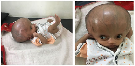
CASE REPORT A term female infant of 27 days, 3rd issue of 2 degree consanguineous marriage born with a big head which was noticed in-utero at 28 weeks of life during routine procedure of the antenatal USG screening. There was no history of recurrent abortion, fever with rash or symptoms suggestive of TORCH infection during first trimester of pregnancy. Baby was delivered by planned lower section caesarean section in view of big size of the baby and mother history of previous two caesarean section. Baby cried immediately after birth, birth weight was 3.5 kg and was kept in NICU for 4 days for observation. On examination the size of the head was increased , dilated veins were present on the scalp, anterior fontanelle was wide open, all sutures were open, there were no hair, impaired upward gaze (setting sun sign) Anthropometric measurement :Head circumference : observed -45cm (34cm), length observed 50cm (52cm), weight 3.9kg (4 kg) On systemic examination of the central nervous system baby was alert with positive glabellar, rooting, sucking, plantar and palmar grasp and incomplete moro reflex. On examination of other system no abnormality was found. The diagnosis was confirmed after CT Brain (plain) in which there was gross dilatation of bilateral lateral and third ventricle with severing thinning of the cerebral parenchyma. Fourth ventricle appeared normal there was also seen obstruction between the third and the fourth ventricle most probably aqueduct stenosis. Cranial sonography was also done which showed gross hydrocephalus with thinning of the brain parenchyma. The baby was treated on emergency basis by doing Ventriculoperitoneal Shunting by the neurosurgeon in which the CSF fluid was shunted to the peritoneum. Baby was in kept in PICU for 1 day for monitoring of the patient. After some time of the surgery there was CSF leak present from the site of operation which was re-sutured again. Baby was shifted to ward the next day and heart rate, respiratory rate was monitored. In the ward the baby was accepting breast feeding and was on inj. ceftriaxone 200mg, inj. amikacin 75mg, inj. levetriacetam 40mg for seven days and patient was discharged and was followed up.
Figure 1 Figure 2 DISCUSSION The term hydrocephalus is derived from a greek word “Hydros” meaning water and “Cephalus” meaning head. As the name suggests it means excessive accumulation of fluid in the brain. The excessive accumulation of the CSF causes the widening of the spaces in the brain known as ventricles. This widening causes potentially harmful pressure on the tissue of the brain. Hydrocephalus can be of two types congenital or acquired. Congenital hydrocephalus is present at birth and may be caused by either events or influences that occur during fetal development, or genetic abnormalities. Acquired hydrocephalus develops at the time of birth or at some point afterward. Hydrocephalus may result from inherited genetic abnormalities (such as the genetic defect that causes aqueductal stenosis) or developmental disorders (such as those associated with neural tube defects including spina bifida and encephalocele).Other possible causes include complications of premature birth such as intraventricular hemorrhage, diseases such as meningitis, tumors, traumatic head injury, or subarachnoid hemorrhage, which block the exit of CSF from the ventricles to the cisterns or eliminate the passageway for CSF within the cisterns. In infancy, the most obvious indication of hydrocephalus is often a rapid increase in head circumference or an unusually large head size. Other symptoms may include vomiting, sleepiness, irritability, downward deviation of the eyes (also called "sun setting"), and seizures. Hydrocephalus is diagnosed through clinical neurological evaluation and by using cranial imaging techniques such as ultrasonography, CT, MRI, or pressure-monitoring techniques. The treatment of this patient is by surgically inserting the shunt which directs the flow of CSF from CNS to another area of the body where it can be absorbed as the normal circulatory process. A shunt is a flexible plastic tube. The shunt system consists of the shunt, the catheter, the valve. Its one end is placed in the in the ventricle and the other is commonly placed in the abdominal cavity. The valve located along the catheter maintains one flow system and circulation of CSF. The overall prognosis of this neonate was bad because of massive dilatation of ventricular system and thinning of cerebral cortex.
REFERENCES
|
|
 This work is licensed under a Creative Commons Attribution-NonCommercial 4.0 International License.