 Home
HomeOfficial Journals By StatPerson Publication
|
Table of Content - Volume 12 Issue 3 -December 2019
A systemic overview of the central venous catheter positioning in chest radiographs of ICU patients
Siddharthkumar B Parmar1, Dinesh Venkatesan2*, Rahul C Manek2, Shweta Girishkumar Shah3
1Assistant Professor, 2Second Year Resident Doctor,3First Year Resident Doctor, Department of Anaesthesiology, B J Medical College, Ahmedabad. Gujarat, INDIA. Email: drsid25@gmail.com
Abstract Background: Central venous catheter is a routine and basic procedure in intensive care unit, required in patients with unstable hemodynamics for fluid transfusion, inotropic support and parenteral nutrition. Chest radiograph is strongly recommended by various guidelines after central venous catheterization to confirm the course, length and tip of catheter. Objective: The aim was to justify the chest radiograph after central venous catheterization and to find out the incidences of malposition of central venous line in critically ill patients. Methods: Seventy patients, who required central venous catheter in Intensive care unit between September to November 2017 and April to July 2018 were enrolled. Catheterization through right subclavian vein was performed under strict asepsis precaution by the anesthetists. Post-catheterization chest radiographs (anterior – posterior views) were performed, and evaluated for course, length and tip of catheter. Malposition and lung complications were also noted. Results: The mean age of patients was 43.97 ± 18.96 years. There were 50(71.43%) males and 20 (28.57%) females in the study. Total 59 (84.29%) CVC were placed correctly having proper course, adequate length of insertion with vertical tip of CVC and no any malposition or lung complications. Total 11(15.71%) CVC were wrongly placed having either improper course, length, tip, malposition or lung complications. In them 6(8.57%) and 2(2.86%) are having either more or less length of CVC insertion respectively. 5 (7.14%) CVC were having angled tip of CVC. 2 (2.86%) were having malposition CVC. There were no any lung complications seen in our study. Conclusion: There was high incidence of incorrect positioning of right sided subclavian vein catheters in intensive care unit. Chest radiographs were accurate to determine the positioning of central venous catheter. Prompt diagnose and early interventions are necessary to reduce the catheter related complications. Key Words: Central venous catheter, chest radiograph, malposition.
INTRODUCTION Central venous catheter (CVC) insertion is utilized in almost all critical ill patients for various purposes like hemodynamic monitoring, fluid transfusion, inotropic supports and parenteral nutrition.1 Ultrasound, Doppler, fluoroscopy and right atrial electrocardiography have been used during CVC insertion but they are not readily available everybody. Chest radiograph after CVC insertion is recommended to determine the correct positioning of CVC.2 It is recommended that the tip of the CVC should be just above the junction of the superior vena cava (SVC) and right atrium at the level of carina. 3,4,5,6. Despite of proper technique, the course, length and tip of CVP may not terminate at desired level leading to incorrect positioning causing lethal complications. The incidence of malposition of CVP are 30% in initial chest radiographs.7 Prompt diagnose and early intervention is essential to reduce CVP related complications. So, the study was carried out to determine the incidence of malposition and complications of CVC using chest radiograph in critically ill patients.
METHODS This was a prospective, descriptive study was carried out after ethical committee approval and written informed consent. Total 70 patients of aged 20 to 70 years old who required CVC during Intensive Care Unit (ICU) stay were included in the study. Patients with altered coagulation, arrhythmia, pacemaker, local site infection were excluded in this study. All patients were monitored for Electrocardiogram, pulse oximetry and noninvasive blood pressure every 5 minute throughout the CVC insertion. All CVP were placed at right subclavian vein by the resident doctors under direct supervision of consultant anaesthesiologist with strict aseptic precautions by blind technique. Chest Radiograph anterior-posterior view was done at end of CVC insertion. Chest radiograph were assessed for course, length and tip of CVP. CVC acquiring normal course of right subclavian vein and tip of CVC be positioned vertically not angled at the level of mid-lower SVC to cavoatrial junction is considered as correct positioned CVC. Proximal or distal to the ideal location of CVC’s tip was considered as incorrect. If CVC don’t acquire normal course of right subclavian vein with kinking, coiling, knotting, fracture, was considered as improper course. Distal end of CVC should lie vertically along the wall of right subclavian vein in chest radiograph and if angled with impingement with the lateral wall of the SVC (>40°), was considered as incorrect. Malposition was defined as CVC entering into any vein other than right subclavian vein. Any lung complications like pneumothorax, hydrothorax, hydro-pneumothorax were also noted. Data were expressed as mean ± SD, numbers and percentages.
RESULTS There were 70 patients with a mean age of 43.97 ± 18.96 years, 50(71.43%) were males and females were 20 (28.57%). Total 59 (84.29%) CVC were placed correctly having proper course, adequate length of insertion with vertical tip of CVC and no any malposition or lung complications. Total 11(15.71%) CVC were wrongly placed having either improper course, length, tip, malposition or lung complications. In them 6(8.57%) and 2(2.86%) are having either more or less length of CVC insertion respectively. 5 (7.14%) CVC were having angled tip of CVC. 2 (2.86%) were having malposition CVC. Both malposition were into ipsilateral internal jugular vein only. There were no any lung complications seen in our study. Table 1: PIVOT TABLE OF CVC POSITIONING
Table 2: Length of CVC
Table 3: Tip of CVC
Table 4: Malposition
Table 5: CVC placement
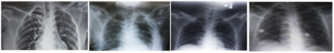
Image 1 Image 2 Image 3 Image 4 Image 1: improper course of CVC – coiling; Image 2: malposition of CVC into Ipsilateral Internal jugular vein 1; Image 3: malposition of CVC into Ipsilateral Internal jugular vein 2; Image 4: More length of CVC into right atrium
DISCUSSION Initial chest radiographs after central venous line show about 30% cases of malposition7. Inaccuracy of methods, anatomical variation, patient and operator variation are the main reasons for the malposition of central venous catheter.8 Vein branches, tortuosity, angulations and stenosis contributes misplacements of central venous catheter. 8 Guide wire placement is also an important step in central line placement, 9, 10 as if it unexpectedly kink and enter into other branch or even run out of the veins, it can cause malposition. Excessive pressure while inserting guide wire can cause malposition. Author believes that resistance to pass guide wire is an important sign of malposition or migration of central venous catheters. Literature3, 4, 5, 6 recommend that CVC should acquire normal course of central vein and tip of CVC should be positioned vertically not angled at the level of mid-lower SVC to cavoatrial junction. Malposition may increase the risk of the vessel wall and cardiac perforation, cardiac tamponade, thrombophlebitis and arrhythmias. CVC can be misplaced into internal mammary vein11, vertebral vein12,13 and azygos vein.14 If CVC abuts the venous wall then it can be migrated into extradural space15, pericardium16, pleural space16, mediastinum16, 17 and thoracic duct.18,19,20 Generally, malposition of CVC results into malfunction early. We believe that malposition of CVC must be promptly diagnosed and early interventions must be done to prevent disasters. This study had demonstrated a high proportional of CVC tips (15.71%) were incorrectly placed. This was slightly lower in a large prospective study21 of 1794 CVC in which they found 6.7% malposition. They defined malposition as CVC tip placement in a vein other than SVC or right atrium, impingement with lateral wall of the SVC (40 °) and arterial cannulation. They inserted CVC via the left internal jugular vein (12%) followed by right subclavian (9.3%), left subclavian (7.3%) and right internal jugular (4.3%). They concluded that even experienced operators cause a considerable number of early mechanical complications and malposition. In this study, a high proportional 6(8.57%) of CVC were placed below the ideal point of insertion. We attributed this to the use of 20 cm CVC in this study. Though we have estimated the depth of insertion by taken a measurement from the site of needle insertion to 3rd/4th rib, still malposition occurred which may be due to individual patient and operator variability during CVC insertion. 2(2.86%) of CVC were placed proximally to superior vena cava which may be due to operator and patient variability or wrong measurement. Placing the CVC tip in a vessel other than the SVC increases the risks of erosion or perforation of vessel walls, local venous thrombosis, catheter dysfunction, and cranial retrograde injection.22 In present study, 5 (7.14%) CVC were having angled tip of CVC and 2 (2.86%) were having malposition CVC. Both malposition were into ipsilateral internal jugular vein only. There were no any lung complications seen in our study. In contrast to our results, the study23 comparing right vs. left subclavian catheterizations in 193 patients undergoing coronary artery bypass graft surgery found malposition of the catheter tip on the right side (9.6%) was significantly more than the left side (0%). In ten patients, catheter tip was located in the ipsilateral internal jugular vein, and in one, in the contralateral subclavian vein. Pneumothorax occurred in 5 patients (4.3%) with right side catheterization only. We used subclavian route for central venous catheter as it is considered to be preferred site to minimize infection risk rather than a jugular or a femoral site.24,25,26,27 Right side was chosen as anatomy of vein and design of catheter facilitate proper insertion24. However, left sided subclavian route will definitely increase the malposition and complications24. Study limitation:
There was high incidence of incorrect positioning of right sided subclavian vein catheters in intensive care unit. Chest radiographs were accurate to determine the positioning of central venous catheter. Prompt diagnose and early interventions are necessary to reduce the catheter related complications.
REFERENCES
|
|
 This work is licensed under a Creative Commons Attribution-NonCommercial 4.0 International License.