 Home
HomeOfficial Journals By StatPerson Publication
|
Table of Content - Volume 12 Issue 3 -December 2019
Rajashri K Virshid1, Unmesh Bedekar2*, Rahul Thorat3
1Associate Professor, 2Junior Resident III, 3Senior Resident, Department of Anaesthesiology, Government Medical College, Aurangabad, Maharashtra, INDIA. Email: rkvirshid@gmail.com, bedekaru@gmail.com, rthorat70@gmail.com
Abstract Background: Juvenile Rheumatoid Arthritis or Still’s disease is a autoimmune disease of childhood. It is a polyarticular disease involving temporomandibular joint giving rise to typical bird facies. The facial deformity pose a challenge to anaesthesiologist because of difficulty in mask ventilation and endotracheal intubation. Case Report: A 25-year-old male of 40 kg weight and known case of juvenile rheumatoid arthritis with micrognathia and malocclusion posted for mandibular distraction surgery. Airway examination revealed difficult mask ventilation and difficult endotracheal intubation. As fibreoptic bronchoscope was not available and there was difficulty in negotiating the south pole tube into trachea, we switched over to retrograde intubation technique and successfully intubated the patient. Retrograde intubation takes up to 4 minutes to accomplish, so it is not useful in a critical emergency airway situation. Conclusion: Retrograde intubation technique is useful in achieving endotracheal intubation in a patient of Juvenile Rheumatoid arthritis with micrognathia. Key Words: Retrograde Intubation, Juvenile Rheumatoid arthritis, micrognathia.
INTRODUCTION Juvenile Rheumatoid Arthritis or Still’s disease is an autoimmune disease of childhood with incidence of 0.4-0.8 per 1,00,000. It is a polyarticular disease with systemic involvement. Most of the time, the temporomandibular joint gets involved and the growth of mandibular condyle stops leading to underdevelopment of mandible i.e. Micrognathia. Hypertrophy of maxilla contributes to deformity of face that is bird face, dental malocclusion. In addition, mouth opening is restricted and there is larger tongue relative to mandible 1. These patients when they cross adolescence are posted for mandibular distraction surgery to decrease the malocclusion and to correct the facial deformity.2 These patients are difficult for mask ventilation and difficult for endotracheal intubation because of the above factors.3 Here we report a case of JRA with micrognathia in which airway was secured with retrograde intubation technique.
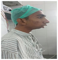
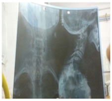
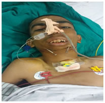
CASE REPORT A 25-year male, weighing 40 kg with diagnosis of Juvenile Rheumatoid Arthritis posted for Mandibular Distraction Surgery. There was history of polyarthritis in childhood at age of 7 years, with stoppage of growth of lower face, malocclusion, difficulty in swallowing solid foods and history of snoring. General examination revealed a small constitution, micrognathia (bird face), malocclusion and maxillary hyperplasia. Airway examination revealed restricted mouth opening with inter-incisor gap of one finger, relatively large tongue, prominent upper incisor, inability to protrude tongue and lower jaw, decreased sterno-mental [11 cm], thyromental [4 cm] and hyo-mental [2 cm] distance, patency of nostrils present. The patient had buck teeth. The upper lip bite test of the patient was positive. The facial profile was complex with incompetent lips. Flexion and extension of head was within normal limits. Systemic examination was insignificant and biochemical investigation was within normal limits. Radiological examination of lateral face X-Ray showed more rostrally placed angle of mandible and more caudally placed hyoid bone .The above parameters suggested difficult mask ventilation and difficult tracheal intubation. Cricothyrotomy set was kept ready for emergency. In view of anticipated difficult mask ventilation and difficult endotracheal intubation, we planned for awake blind nasal intubation with backup plan of retrograde intubation and last resort of tracheostomy as there was unavailability of fibreoptic bronchoscope which is considered gold standard in such cases. On the day of surgery with monitors attached, IV line was set up, patient was premedicated with injection glycopyrrolate 0.02mg/kg. Airway preparation started with instillation of oxymetazoline nasal drops in both nostrils followed by nebulisation with 5 cc of 2 % lignocaine for 5 minutes, gauze soaked in lignocaine was kept in both nostrils for 3 minutes, superior laryngeal nerve block was given with 2 % lignocaine 4 cc and transtracheal block with 2 % lignocaine 2 cc. Premedication with injection Dexmedetomidine 0.5 ug/kg bolus followed by infusion of 0.5 ug/kg/hr. Ivory north pole endotracheal tube no 7 lubricated with 4% lignocaine and inserted through nostrils with manoeuvres done to achieve endotracheal intubation but there was difficulty in negotiating the tube through larynx and repeated oesophageal intubation was occurring; therefore the procedure was abandoned .Gag reflex was strong and repeated lignocaine spraying was done. But we were unable to intubate. According to backup, retrograde intubation procedure was initiated. 2 % lignocaine 1 cc was injected subcutaneously in the centre of cricothyroid membrane .The membrane was punctured with 18 G epidural needle and 18 G epidural catheter was passed through it in cephalad direction .The catheter came out of the mouth .Ryles tube no.14 was passed through nostril and brought out of the mouth .Epidural catheter was railroaded through the Ryles tube and brought out through the nostril. The Ryle’s tube was removed. The catheter was threaded through the Murphy’s eye inside the lumen of ETT no .6.5 and brought cut from proximal end of ETT. Both ends of catheter were given adequate tension. ETT was manipulated through the laryngeal inlet into the trachea. The position was confirmed by EtCO2 graph. Epidural catheter was kept in situ intraoperatively. Patient was induced with Injection Propofol 2.5 mg/kg, patient was maintained on oxygen 50% and nitrous oxide 50% and isoflurane and injection vecuronium 0.1 mg/ kg. At the end of surgery, patient was reversed with Injection Neostigmine 0.5mg/kg and Injection Glycopyrrolate 8 ug/kg done. Patient was shifted to post-anaesthesia care unit for 24 hours observation. Patient was extubated after 24 hours but epidural catheter was kept in situ for further 24 hours. Injection hydrocortisone 100 mg IV and Injection Dexamethasone 8 mg IV were given. NBM was released after 6 hours and oral sips were started. Undue pressure at the head end was avoided and proper 30-degree head elevation was given.
DISCUSSION In our patient, the history, general examination, airway examination and radiological finding suggested difficult mask ventilation and intubation according to parameters given in review article. Fibreoptic intubation is the gold standard for securing airway in such cases. Previous case reports have used Fibreoptic intubation alone 4,5,6 or in conjunction with Laryngeal Mask Airway7. But unavailability of fibreoptic bronchoscope precluded us from using it .Use of ILMA and video laryngoscopy has been recommended in such cases8.That too was not available at our place. Blind nasal intubation has been used in cases of temporomandibular joint ankylosis8 and also, in case of high mallampatti grade9. The difficulty we faced with blind nasal intubation can be due to the strong gag reflex which could have been obtunded by glossopharyngeal block10, but due to anatomical distension of face, we did not do it. Also, the rostrally placed mandibular angle and caudally placed hyoid bone caused the tongue to occupy more of hypopharynx than oropharynx and this must have added to our difficulty. Also, cricoarytenoid arthritis leading to fixation of vocal cords is common in JRA11 which may have added to difficulty in achieving endotracheal placement by nasal route. Retrograde intubation is indicated in cases of micrognathia, large tongue, limited mouth opening with protruding tongue, failed blind intubation and failed fibreoptic intubation12 and also in case of difficult oral intubation with direct laryngoscopy13. We could easily achieve endotracheal intubation with this technique and every anaesthesiologist should be well-versed with this retrograde intubation technique.14 Injection Glycopyrrolate as an anti-sialagogue agent ensures dry field and allows local anaesthetics to act on mucosal surface in appropriate concentration. Oxymetazoline helps in nasal decongestion of highly vascular mucosa of nose and nasopharynx. Injection Dexmedetomidine achieves sedation without affecting ventilation and also has analgesic action15. Topical local anaesthesia and regional airway blocks facilitate awake intubation10,15 .We kept the epidural catheter in situ intraoperatively in case there is accidental withdrawing of tube through laryngeal inlet as the tip of ETT is just below the vocal cords while in routine ETT intubation, the tube is 4-6 cm beneath the cords. The epidural catheter was kept in situ post-operatively after extubation for 24 hours just in case there is oedema formation and need of reintubation arises.
Preoperative Postoperative
CONCLUSION Juvenile rheumatoid arthritis with micrognathia can be safely intubated with retrograde intubation if other techniques of intubation fail or when fibreoptic bronchoscope is not available.
REFERENCES
|
|
 This work is licensed under a Creative Commons Attribution-NonCommercial 4.0 International License.