 Home
Home
|
Table of Content Volume 16 Issue 2 - November 2020
Use of non-contrast computed tomography for evaluation of adrenal volume in a tertiary care centre and to establish a normative value in Indian population
Vishwaprem Raj D R
Assistant Professor, Department of Radiodiagnosis, Sapthagiri Institute of Medical Sciences & Research Centre, Bengaluru, INDIA. Email: vishwapremrajdr@gmail.com
Abstract Background: Size and shape of the adrenal glands varies in different individuals. Adrenal volume is a good indicator to make comparison compared to other parameters. Adrenal gland shape and volume have been described in a very few published articles, mostly in western and Chinese populations. No studies regarding adrenal volume have been published till date regarding Indian population. Purpose: To find volume of adrenal glands in adult patients reported for CT abdomen without any clinical evidence of diseases related to adrenals. Materials and Methods: 100 patients (50 males and 50 females) without any clinical evidence of diseases affecting adrenal glands and with suspected renal or ureteric colic, referred to department of radio-diagnosis – Sapthagiri Institute of Medical Sciences and Research for NCCT evaluation of abdomen. Results: Mean volume of right adrenal gland is 3.32cc (SD 0.454) in males and 3. 32cc (SD 0.454) in females and the mean value of the left adrenal gland is 3.51cc (SD 0.432) in males and 3.23 cc (SD 0.426) in females. Conclusion: There is no significant difference in the mean right and total adrenal gland volumes between males and females. However, there is statistically significant difference in volumes of the left adrenal gland, being higher in males. No significant change seen between the males and females in respect to shape of right and left adrenal glands. Key Words: Normal adrenal volume, CT adrenal volume, Normal adrenal shape.
INTRODUCTION Adrenal glands are vital endocrine glands, which play a crucial role in many metabolic processes. Adrenal glands are tiny structures situated in the extra-peritoneal peri-renal space on both sides1. Adrenal glands are affected by wide variety of diseases which lead to morphological changes of the gland2. Adrenal gland volume can also be modified by several systemic pathological conditions including depression and shock3. Adrenal volume measurement is now possible with the advent of multi-detector CT4. Till date, very few studies have quantified adrenal gland volume. CT volume of adrenal glands in Indian population have so far been not published. The purpose of this article is to measure volume normal adrenal glands in adult Indian population, to evaluate relationship of volume of adrenal glands with age and to assess the correlation between these factors. CT in evaluation of adrenal morphology: MDCT is a primary imaging tool for adrenal disease5. Multi-detector CT is capable of the rapid image acquisition that is required for the routine use of multi-phasic contrast-enhanced examination6. It also provides thin-section imaging of the adrenal glands and is capable of obtaining volumetric data sets that can be used with post-image processing techniques. CT anatomy: The adrenal gland visualization on CT depends upon the presence of retroperitoneal fat. In-adequate suspension of respiration, lack of retroperitoneal fat and streak artifacts can contribute to non-visualization of normal adrenal in some individuals7. Normal shape of adrenals is determined on the section where the gland is best seen. In most cases the right gland appears linear or slightly curvilinear. The left gland has more of a V or a Y shape, but occasionally appears more square or triangular. The length of each adrenal gland is estimated by counting the number of transverse cross sections on which each is visualized. The normal adrenal gland length is 2-4cm5. The width of each gland is determined on the section in which the adrenal appears largest. About 75% of the right adrenal glands and 80% of the left glands have maximal width of 2.0-2.5cm5. The thickness is often less than 8mm thick or less.
MATERIALS AND METHODS 100 patients (50 males and 50 females) without any evidence of diseases affecting adrenal glands (no clinical, dermatological or biochemical parameters suggestive of adrenal pathologies) and with suspected history of renal or ureteric colic, referred to department of radio-diagnosis – SIMSandRC for NCCT evaluation of abdomen. Patients were counselled and only those patients willing to participate in the study were included. After taking informed consent, plain abdominal scan was obtained with Toshiba Activion 16 slice multi-detector CT; 5mm slice thickness to rule out any obvious intra-abdominal pathology. Adrenals were then imaged employing 1.25mm sections and each adrenal gland was traced in all axial slices. Volume of the adrenal gland was calculated by summing areas for all relevant sections and multiplying by a factor of 1.25, as slice thickness of images was 1.25mm. The data thus collected was compiled in Microsoft Office Excel 2013 format. Original data is presented as means and standard deviations. The comparison of adrenal gland volumes between males and females was done by a "Two tailed Paired T Test" and p value was calculated.
RESULTS The mean age of the study population is 28 years. The mean age of male patients is 27 years and females is 31 years. There were a total of 55 cases in the group of 21 to 30 years, including 23 females and 32 males. In the age group of 31 to 40 years, there were 18 male and 27 female patients. The mean adrenal volume of the study population is 3.32cc (SD 0.436 cc) for right adrenal gland and 3.37cc (SD 0.442 cc) for left adrenal gland. The mean total adrenal gland volume is 6.69cc (SD 0.79 cc). The mean left adrenal gland volume is marginally more than the right adrenal gland, however the difference is insignificant (0.05cc i.e. 1.5%). On further analysis, in females, the right adrenal gland volume was found to be marginally larger (0.23cc i.e. 7.3%) in the age group of 3rd decade (21 to 30 years) in comparison with age group of 4th decade (31 to 40 years). However the left adrenal gland was smaller in the 3rd decade age group as compared to 4th decade, by 0. 22cc (3%). The total adrenal gland volume was larger in the 3rd decade age group, by 0.17cc. In males, the right adrenal gland volume was found to be smaller (0.16cc i.e. 4.8%) in the age group of 3rd decade (21 to 30 years) in comparison with age group of 4th decade (31 to 40 years). Left adrenal gland was again smaller in the 3rd decade age group, by 0.22cc. The total adrenal gland volume was significantly larger in 4th decade age group in comparison with 3rd decade group by 0.38cc (11%). (Table 1).
Table 1: Analysis of adrenal gland volumes in males and females
There is no significant difference in the mean right and total adrenal gland volumes between males and females (p values 0.76 and 0.137 respectively for right and total gland volumes). However there is a statistically significant difference in the volumes of left adrenal glands (p value— 0.026), being larger in males. (Table 2).
Table 2: Difference in volume between males and females
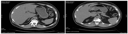
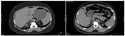
The most common shape of the right adrenal gland, encountered in the study is linear shape (90%). Curvilinear shape (7%) was second most common. The most common shape of the left adrenal gland in this study is V shaped (78%), followed by Y shaped (16%) and linear (4%). It is also observed that the more common shape of right adrenal gland in male and female is leaner and most common shape of Left adrenal is V shape. No difference in shapes of adrenal glands between males and females was observed. Figure 1: Manual tracing of adrenal gland volume 2a: 2b Figure 2- 2a: NCCT - Axial section showing Y shaped left adrenal gland; 2b: NCCT – Axial section, showing linear right adrenal gland and triangular left adrenal gland
DISCUSSION The adrenal gland volume has significant clinical importance in several crucial conditions3, 8,9. Mean volume of the adrenals varies with different investigators, with the mean value ranging from 3.5 to 11.4 cc. All previous studies were conducted in western and Chinese and Western populations4, 8 and to best of the knowledge of the authors, there is no published article on Indian population. This study was therefore conducted to determine the range of normal adrenal gland volume in Indian adult subjects. In this study, patients coming to the institution from various states of India for medical care were evaluated, as our institute is strategically located to cater to both north and south Indian population. As such, the sample taken can be fairly considered as representative of Indian population. The mean total volume adrenal gland in this study was 6.9cc, which was in accordance with the results of previous studies8, 11. The volume of normal Right adrenal gland and left adrenal gland as observed by Rubin et al.4, involving Western population, was 3.62cc and 3.73 cc respectively. Amsterdam JD et al. in 19878, measured the volume of right adrenal gland and left adrenal glands and found them to be 3.9±2.1 cc, 4.1±2.8cc respectively. Again, the left adrenal is marginally larger than the right adrenal gland in terms of volume. As per a study by Geraghty EM et al., the volume of adrenal gland range between 3 to 4.4cc which is in concordance with the present study12. Another study by Nougaret S et al.., found the total volume of adrenal glands to be 7.2±2cc9. In another study done by Xuan Wang et al.13, involving Chinese population, the mean right adrenal gland volume was 4.26cc and left adrenal gland volume was 4.23cc. The mean total adrenal gland volume was 8.5cc which is larger than the mean total volume found in the present study population. In contrast to previous studies of Rubin and Phillips4, the relative contribution of gender to adrenal volume is not significant, except left adrenal gland volume, which show statistically significant larger volume in males as compared to females. No consistent correlation observed between adrenal volume and age as also observed by Wang X et al.13. Adrenal gland shape: The shape of the right and left adrenal gland varies because of its anatomical location and adjacent anatomical relation to other organs. Nevertheless, linear measurements are affected by variety of configurations of the adrenal gland. Assessing adrenal volume, thereby is an accurate way which overcomes the effect of gland morphology on measurement. As per studies done by Vincent et al., the most common shape of the adrenal gland are linear / curvilinear on right side and V or Y shape on the left side, which are similar to that found in our study.
LIMITATIONS Manual tracing of the adrenals was done. Variations while tracing the adrenal gland are unavoidable. Use of semi-automated organ segmentation techniques can standardize volumetry to an extent.
CONCLUSION This study has defined the volume distribution and shape of normal adrenal glands in adults. These measurements provide a baseline for establishing adrenal normality or enlargement on CT examination of the adrenal glands, which are useful to facilitate accurate diagnosis in clinical or research setting. By this study, we conclude that the mean volume of adrenal in Indian population slightly differ from the Chinese and Western population and therefore we requires more studies to standardize the adrenal gland volume precisely. Besides, the most common shape of right adrenal gland is curvilinear and left adrenal gland is V shape in Indian population. CT is the modality of choice to assess adrenal gland volumes.
SUMMARY The mean volume of Right adrenal gland is 3.32cc (SD 0.454) in males and 3.32cc (SD 0.454) in females and the mean volume of the Left adrenal gland is 3.52cc (SD 0.432) in males and 3.23cc (0.426) in females. There is no significant difference in the mean right and total adrenal gland volumes between males and females (p values 0.76 and 0.137 respectively for right and total gland volumes). However there is a statistically significant difference in the volumes of left adrenal glands (p value= 0.026), being larger in males.
REFERENCES
Policy for Articles with Open Access: Authors who publish with MedPulse International Journal of Radiology (Print ISSN: 2579-0927) (Online ISSN: 2636-4689) agree to the following terms: Authors retain copyright and grant the journal right of first publication with the work simultaneously licensed under a Creative Commons Attribution License that allows others to share the work with an acknowledgement of the work's authorship and initial publication in this journal. Authors are permitted and encouraged to post links to their work online (e.g., in institutional repositories or on their website) prior to and during the submission process, as it can lead to productive exchanges, as well as earlier and greater citation of published work.
Height, IVDL-Intervertebral Disc Length
Policy for Articles with Open Access
Authors who publish with MedPulse International Journal of Radiology(Print ISSN: 2579-0927) (Online ISSN: 2636 - 4689) agree to the following terms: Authors retain copyright and grant the journal right of first publication with the work simultaneously licensed under a Creative Commons Attribution License that allows others to share the work with an acknowledgement of the work's authorship and initial publication in this journal. Authors are permitted and encouraged to post links to their work online (e.g., in institutional repositories or on their website) prior to and during the submission process, as it can lead to productive exchanges, as well as earlier and greater citation of published work. |
|
|||||||||||||||||||||||||||||||||||||||||||||||||||||||||||||||||||||||||||||||||||||||||||||||||||||
 This work is licensed under a Creative Commons Attribution-NonCommercial 4.0 International License.