 Home
Home
|
Table of Content Volume 16 Issue 3 - December 2020
Study of duplex colour doppler imaging of carotid arteries in patients with peripheral vascular disease in Telangana population
Balaji Patel Kola1, Abhijith Kumar Kotte2*
1Associate Professor, 2Assistant Professor, Department of Radiology, Apollo Hospitals, Apollo Institute of Medical Sciences and Research, Jubilee Hills, Hyderabad, Telangana State, INDIA. Email: drbalaji777@gmail.com
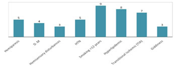
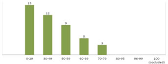
Abstract Background: Atherosclerosis of carotid arteries associated with peripheral vascular diseases like claudication, TAO, TAI, Hemisensory disturbances etc. Method: 44 patients aged between 30 to 75 years were exposed to Duplex colour Doppler, USG, Angiography. High-frequency linear array transducer (5, 7 or 10 MHz) was used. Carotid artery walls and their velocities were studied in the neck on both sides in longitudinal and transverse planes different measurements were taken to grade the stenosis. Results: Clinical manifestation were – 5 (11.3%) had hemiparesis, 4 (9%) had DM, 3 (6.8%) had hemisensory disturbances, 5 (11.3%) had HTN, 9 (20.9%) had smoking history >12 years , 8 (18.1%) had hyperlipidemia, 7 (15.9) had TIA, 3 (6.8%) had giddiness. The grades of ICA stenosis with percentage was, 15 (33.3%) patients had 0- 29% stenosis, 12 (27.2%) had 30-49%, - 9 (20.4%) patients had 50-59%, 5 (11.3%) had 60-69% stenosis, 3 (11.3%) had 70-79% of stenosis. Conclusion: As there is a close association between carotid artery disease with peripheral vascular disease. Duplex ultrasound is accurate, safe and economic. Keywords: Stenosis, TIA-Transient Ischemic attack, TAO – Thrombo angiitis obliterans, DM – Diabetic Mellitus, PVD – Peripheral Vascular disease.
INTRODUCTION Cerebral Ischemic stroke is a major cause of death. Atherosclerosis is an intra and extracranial carotid vessel pathology, leading to cerebral infarction accounting for 80% of strokes. It has conclusively proven that the risk of major stroke is higher in the first 3 months of transient ischemic attack (TIA)1 and It was also reported that 20% or more strokes have been heralded by TIA2. The highest risk of large artery stroke appears to be among patients with the highest degree of carotid stenosis, history of DM, HTN, atherosclerosis, chronic smokers3. Duplex colour Doppler imaging evaluation of carotid arteries and endarterectomy in patients having PVD will be an easy method to predict the severe consequences of stroke4 death due to cerebral infarction or haemorrhage.
MATERIAL AND METHOD 44 (forty-four) patients aged between 30 to 75 years referred by the medicine department of Apollo Hospital were studied at the Radiology department of Apollo Hospital, Hyderabad-500096, Telangana. Inclusion: Patients having peripheral vascular symptoms of Cerebro – basilar insufficiency symptoms. History of transient ischemic attack with Diabetes mellitus, TAO were selected for the study. Exclusive Criteria: Patients already under treatment for epilepsy, malignancy, psychosis, HIV were excluded from the study. Method: All of them were diagnosed as having peripheral vascular disease, based on clinical history and examination, Duplex colour doppler ultrasonography, Angiography. USG performed with ultrasound Machine GE voluson PS Machine. High-frequency Linear-array transducer (5, 7 or 10 MHz) was used. Carotid arterial walls and their velocities were studied in the neck on both sides in the longitudinal and transverse planes. Three measurements
The duration of the study was about 2 years (March-2018 to April-2020) Statistical analysis: The clinical manifestations, grades of stenosis in patients were classified with percentage. The statistical analysis was done in SPSS software. The ratio of male and females was 2:1.
OBSERVATION AND RESULTSTable-1: Clinical manifestation of patients studied 5 (11.3%) had hemiparesis, 4 (9%0 had DM, 3 (6.8%) had Hemi sensory disturbances, 5 (11.3%) had HTN, 9 (20.4%) patients had the habit of smoking more than 12 years, 8 (18.1%) had hyperlipidemia, 7 (15.9%) had TIA 3 (6.8%) had giddiness. Table-2: Grades of ICA stenosis with percentage in patients – 15 (13.3%) patients had 0-29% 12 (27.2%) had 30-49% , 9(20.4%) had 50-59%,5 (91.3%) had 60-69% of stenosis , 3 (6.81%) had 70-79 of stenosis.
Table 1: Clinical manifestation of patients presented (No. of patients: 44)
Table 1: Clinical manifestation of patients presented
Table 2: Grades of Internal Carotid Artery stenosis in patients presented with percentage
Table 2: Grades of internal carotid artery stenosis in patients presented with percentage
DISCUSSIONThe present study of duplex colour Doppler imaging of carotid arteries with peripheral vascular disease in Telangana Population. The clinical manifestations were 5 (11.3%) had hemiparesis, 4 (9%) had DM, 3 (6.8%)had Hemisensory disturbances, 5 (11.3%) had HTN, 9 (20.4%) with smoking history > 12 years, 8 (18.1%) with Hyperlipidemia , 7 (15.9%) had TIA, 3 (6.8%) had giddiness (Table-1). The grades of ICA stenosis were – 5 (33.3%) patients had 0-29% , 12 (27.2%) had 30-40% stenosis, 9 (20.4%) had 50-59%, 5 (11.3%) had 60-69% of stenosis, 3 (6.8%0 had 70-79% of stenosis (Table-2). These findings were more or less in agreement with previous studies5,6,7. It is reported by (NASCET) North American Symptomatic Endarterectomy Trial and (ECST) European carotid surgery Trial, that evaluation of degrees of carotid stenosis will help to reduce with help to the surgical risk(8), hence there was a significant reduction in the stroke in the patients having carotid stenosis and a surgical perioperative morbidity/ mortality was reduced to 2%9. It is an established fact that the benefit of carotid endarterectomy depends on optimal surgical expertise and careful patient’s selection. PVD is a known risk factor to cause a stroke. It has increased 2 to 5 fold risk due to hypercholesterolemia and overweight of patients10 Apart from this smoking, HTN, DM have a significant correlation to stroke due to elevation in the lipids (cholesterolemia) which mainly involves coronary artery diseases (CAD). In the present study up to 70-79% of stenosis was observed but previous studies have observed complete (100% occlusion (stenosis) of ICA(11). In such cases absence of pulsation, lumen filled echogenic material, subnormal vessel size and absence of Doppler flow signals or weak Doppler signals were observed.
SUMMARY AND CONCLUSIONThe present study duplex of colour Doppler imaging carotid arteries in patients with PVD in the Telangana population has proved that there is a close relation between carotid artery disease and PVD. Duplex ultrasound is an accurate safe and economic imaging technique of screening high-risk patients who had already been investigated for primary disease. Such cheaper duplex ultrasound is most suitable for developing countries like India. This research paper was approved by the Ethical committee of Apollo Hospital, Apollo Institute of Medical Sciences and research, Jubilee Hills, Hyderabad - 500096. REFERENCES
Policy for Articles with Open Access: Authors who publish with MedPulse International Journal of Radiology (Print ISSN: 2579-0927) (Online ISSN: 2636-4689) agree to the following terms: Authors retain copyright and grant the journal right of first publication with the work simultaneously licensed under a Creative Commons Attribution License that allows others to share the work with an acknowledgement of the work's authorship and initial publication in this journal. Authors are permitted and encouraged to post links to their work online (e.g., in institutional repositories or on their website) prior to and during the submission process, as it can lead to productive exchanges, as well as earlier and greater citation of published work.
Height, IVDL-Intervertebral Disc Length
Policy for Articles with Open Access
Authors who publish with MedPulse International Journal of Radiology(Print ISSN: 2579-0927) (Online ISSN: 2636 - 4689) agree to the following terms: Authors retain copyright and grant the journal right of first publication with the work simultaneously licensed under a Creative Commons Attribution License that allows others to share the work with an acknowledgement of the work's authorship and initial publication in this journal. Authors are permitted and encouraged to post links to their work online (e.g., in institutional repositories or on their website) prior to and during the submission process, as it can lead to productive exchanges, as well as earlier and greater citation of published work. |
|
 This work is licensed under a Creative Commons Attribution-NonCommercial 4.0 International License.